. 2023 Feb;8(2):109-120.
doi: 10.1016/j.jacbts.2022.09.001. Epub 2022 Sep 14.

Human Cardiac Pericytes Are Susceptible to SARS-CoV-2 Infection

Affiliations

Human Cardiac Pericytes Are Susceptible to SARS-CoV-2 Infection

Brittany D Brumback et al. JACC Basic Transl Sci. 2023 Feb.

Abstract

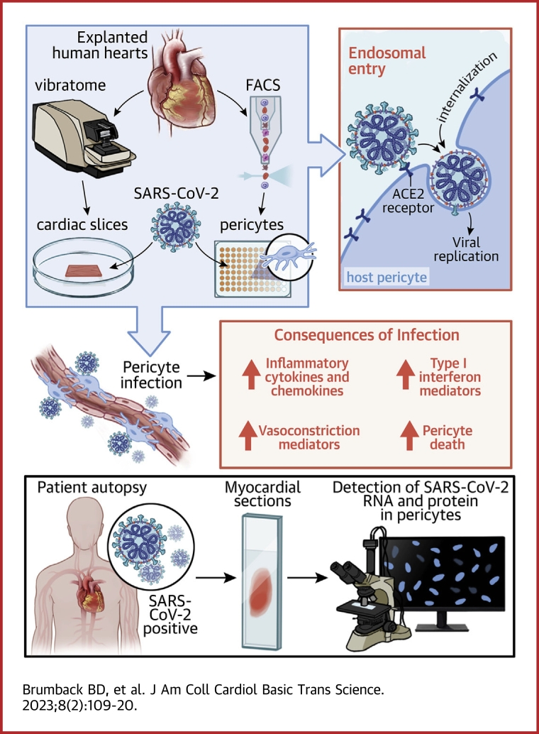
COVID-19 is associated with serious cardiovascular complications, with incompletely understood mechanism(s). Pericytes have key functions in supporting endothelial cells and maintaining vascular integrity. We demonstrate that human cardiac pericytes are permissive to SARS-CoV-2 infection in organotypic slice and primary cell cultures. Viral entry into pericytes is mediated by endosomal proteases, and infection leads to up-regulation of inflammatory markers, vasoactive mediators, and nuclear factor kappa-B-dependent cell death. Furthermore, we present evidence of cardiac pericyte infection in COVID-19 myocarditis patients. These data demonstrate that human cardiac pericytes are susceptible to SARS-CoV-2 infection and suggest a role for pericyte infection in COVID-19.

Keywords: ACE2, angiotensin-converting enzyme 2; COVID-19; IFN, interferon; MOI, multiplicity of infection; PDGFRβ, platelet-derived growth factor receptor beta; SARS-CoV-2; cardiovascular disease; pericytes.


Conflict of interest statement

This research was supported by National Institutes of Health/National Center for Advancing Translational Sciences grant UH3 HL141800-04S1 COVID-19 supplement for an emergency response to COVID-19 (to Dr Rentschler), National Institutes of Health National Heart, Lung, and Blood Institute/National Center for Advancing Translational Sciences UH3 HL 141800 (to Dr Rentschler), and the National Science Foundation GRFP grant DGE-1745038 (to Dr Brumback). The Genome Technology Access Center at the McDonnell Genome Institute at Washington University School of Medicine is partially supported by NCI Cancer Center Support Grant #P30 CA91842 to the Siteman Cancer Center and by ICTS/CTSA Grant# UL1TR002345 from the National Center for Research Resources, a component of the National Institutes of Health, and National Institutes of Health Roadmap for Medical Research. Dr Diamond is supported by the National Institutes of Health (R01 AI157155); is a consultant for Inbios, Vir Biotechnology, Moderna, Immunome, and Senda Biosciences; and has received unrelated sponsored research agreement funding from Moderna, Vir Biotechnology, and Emergent BioSolutions. Dr Lavine is supported by the National Institutes of Health (R01 HL138466, R01 HL139714, R01 HL151078), Leducq Foundation Network (#20CVD02), Burroughs Wellcome Fund (1014782), Children’s Discovery Institute of Washington University and St. Louis Children’s Hospital (CH-II-2015-462, CH-II-2017-628, PM-LI-2019-829), and Foundation of Barnes-Jewish Hospital (8038-88); is a consultant for Implicit Biosciences and Flame Biosciences; is a member of the Medtronic: DT-PAS/APOGEE trial advisory board; and has received funding and unrelated sponsored research agreements from Amgen and Novartis. Dr Rentschler is supported by the National Institutes of Health (R01 HL130212, UH3 HL141800), Burroughs Wellcome Fund (1009884), Foundation of Barnes-Jewish Hospital, and Additional Ventures Cures Collaborative. All other authors have reported that they have no relationships relevant to the contents of this paper to disclose.

Figures

None
Graphical abstract
Figure 1
Figure 1
Detection of SARS-CoV-2 in Pericytes of Inoculated Ventricular Organotypic Slices (A) Representative immunostaining for angiotensin-converting enzyme 2 (ACE2) (green) and platelet-derived growth factor receptor beta (PDGFRꞵ) (red) in human left ventricular tissue (n = 3 independent donor hearts). (B to D) Cardiac slices were inoculated with 5 × 105 focus forming units of SARS-CoV-2 WA1/2020 (approximately MOI of 1) and harvested 48 hours later (n = 3). (B) Representative RNA in situ hybridization of SARS-CoV-2 S sense (S-S) (green) and SARS-CoV-2 ORF1ab antisense (O-AS) (red) at 48 hours postinoculation. (C) Representative RNA in situ hybridization of SARS-CoV-2 S sense (S-S) (green) and pericyte marker PDGFRB sense (red), and (D) immunostaining for the SARS-CoV-2 NP (green) with pericyte marker PDGFRꞵ (red). All scale bars: 10 μm. All images representative from 3 technical slice replicates from each of the 3 independent donor hearts. DAPI = 4′,6-diamidino-2-phenylindole.
Figure 2
Figure 2
SARS-CoV-2 Productively Infects Human Primary Cardiac Pericytes (A) Immunostaining for ACE2 (green) and PDGFRꞵ (red) of cardiac pericytes isolated from left ventricle (n = 3 donors). (B) Representative flow cytometry histogram and quantification of PDGFRꞵ in pericytes (n = 3). (C and D) Focus-forming assay time course of pericyte infection with SARS-CoV-2 WA1/2020 and SARS-CoV-2 mNeonGreen (n = 3) (C) and Alpha (B.1.1.7), Beta (B.1.351), and Delta (B.1.617.2) SARS-CoV-2 variants (representative of n = 2) (D); dotted line indicates limit of detection. (E) Immunostaining for the SARS-CoV-2 nucleocapsid protein (NP) (green) and PDGFRꞵ (red) (n = 3). (F) Representative flow cytometry histogram and quantification of cardiac pericyte infection (mNeonGreen) at day 1 postinoculation with multiplicity of infection (MOI) of 20 (n = 3). (G) Flow cytometry-based cell viability (ZombieViolet+ = dead) time course of inoculated pericytes. Statistical comparison to mock at corresponding time point. (H to J) Pericytes were pretreated with vehicle (unt), 10 μg/mL of human anti-ACE2 antibody (α-hACE2), 100 μmol/L of serine protease inhibitor camostat methylate (camo), 100 μmol/L of cysteine protease inhibitor E-64 (E64), or a combination of camostat methylate and E-64 (camo-E64) for 1 hour, inoculated with mock or SARS-CoV-2 mNeonGreen at MOI of 20 for 20 hours (n = 2). Quantification of infection (mNeonGreen+) (D), pericyte identity (PDGFRꞵ+) (E), and cell viability (Zombie-Violet-) (F). (D to F) Points represent separate wells; statistical comparison to untreated inoculated group. Repeated in 2 donors. 2-way analysis of variance with Dunnet’s post-test: ∗∗P < 0.01; ∗∗∗P < 0.001. Scale bars: 20 μm. Abbreviations as in Figure 1.
Figure 3
Figure 3
SARS-CoV-2 Infection Upregulates Type I IFN Signaling, Inflammatory Pathways, and Vasoactive Genes in Cardiac Pericytes (A to G) RNA sequencing of pericytes at 4 dpi. Cardiac pericytes from 3 donors were inoculated with SARS-CoV-2 mNeonGreen (5 technical replicates/donor) or mock (3 technical replicates/donor). (A) Multidimensional scaling plot with mock (red) and SARS-CoV-2 infected (CoV2) (green) samples; dots represent technical replicates. (B) Volcano plot showing differentially expressed genes between mock and infected samples. Each dot represents a gene, with blue denoting down-regulated genes, black denoting genes nonsignificant changes, and red denoting up-regulated genes. (C) Gene ontology pathway analysis with top 10 up-regulated pathways in infected pericytes. (D to G) Heat maps of selected differentially expressed genes of viral origin (D), pericyte genes involved in inflammatory response (E); response to IFN signaling (F); and vascular development, homeostasis, and function (G). (H to J) Reverse transcription quantitative polymerase chain reaction analyses of genes encoding inflammatory cytokines and chemokines (H), type I interferon signaling pathway components (I), and vasoconstriction and angiogenesis mediators (J); data points denote separate wells (3 technical replicates/donor) with statistical comparison to mock group for each donor; an equal or unequal variance Student's t-test, as determined by an F test for equality of 2 variances: ∗P < 0.05; ∗∗P < 0.01; ∗∗∗P < 0.001, nonsignificant comparisons not shown. GAPDH = glyceraldehyde 3-phosphate dehydrogenase; other abbreviations as in Figure 1.
Figure 4
Figure 4
Detection of SARS-CoV-2 in Pericytes of COVID-19 Cardiac Autopsy Specimens (A) RNA in situ hybridization of SARS-CoV-2 S sense (S-S) (green) with pericyte marker PDGFRB sense (PDGFRB-S) (red) in the perivascular region of autopsy control (n = 1) and COVID-19–positive formalin-fixed paraffin-embedded specimens (n = 2). (B) Immunostaining for the SARS-CoV-2 NP (green) and pericyte marker PDGFRꞵ (red) within the perivascular region of autopsy control (n = 1) and COVID-19–positive formalin-fixed paraffin-embedded specimens (n = 2). Abbreviations as in Figure 1.

Comment in

References

    1. Gupta A., Madhavan M.V., Sehgal K., et al. Extrapulmonary manifestations of COVID-19. Nat Med. 2020;26:1017–1032. - PMC - PubMed
    1. Katsoularis I., Fonseca-Rodríguez O., Farrington P., Lindmark K., Connolly A.-M.F. Risk of acute myocardial infarction and ischaemic stroke following COVID-19 in Sweden: a self-controlled case series and matched cohort study. Lancet. 2021;398:599–607. - PMC - PubMed
    1. Nägele M.P., Haubner B., Tanner F.C., Ruschitzka F., Flammer A.J. Endothelial dysfunction in COVID-19: Current findings and therapeutic implications. Atherosclerosis. 2020;314:58–62. - PMC - PubMed
    1. Sims J.T., Krishnan V., Chang C.-Y., et al. Characterization of the cytokine storm reflects hyperinflammatory endothelial dysfunction in COVID-19. J Allergy Clin Immunol. 2021;147:107–111. - PMC - PubMed
    1. Pellegrini D., Kawakami R., Guagliumi G., et al. Microthrombi as a major cause of cardiac injury in COVID-19: a pathologic study. Circulation. 2021;143:1031–1042. - PubMed