

Compass Fundus-Guided Perimetry in Geographic Atrophy
- PMID: 36124138
- PMCID: PMC9482498
- DOI: 10.1155/2022/1315588
Compass Fundus-Guided Perimetry in Geographic Atrophy
Abstract
Purpose: To evaluate compass (CMP), a recently introduced device that combines scanning ophthalmoscopy, automated perimetry, and eye tracking, for fundus-guided perimetry (microperimetry) with the purpose of correlating perimetric retinal sensitivity (PRS) and retinal geographic atrophy (GA) features.
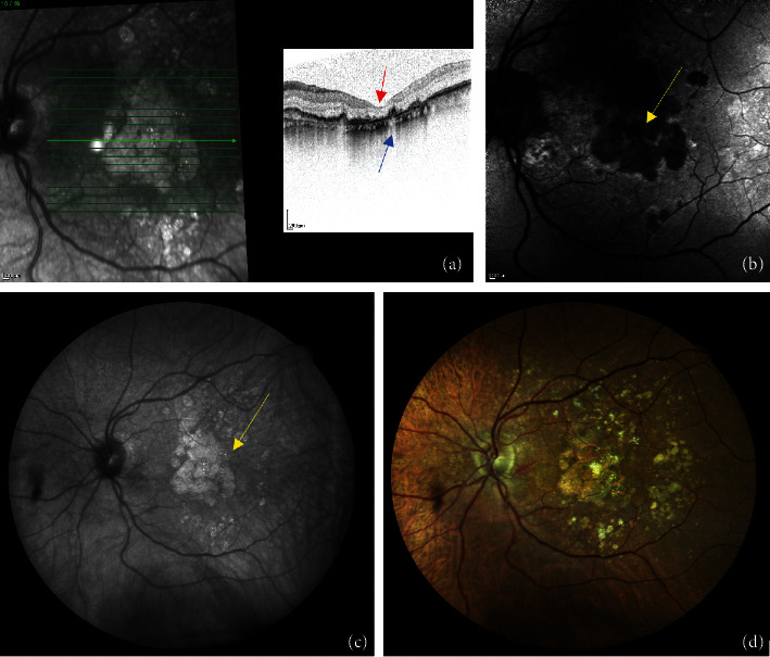
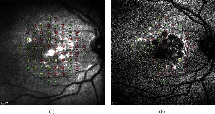
Materials and methods: A retrospective, cross-sectional study was performed in 56 eyes of 43 patients affected by GA. All patients underwent compass 10-2 perimetry, consisting of a full-threshold visual field on fundus photography and an infrared (IR) image of the central 30° of the retina. Data were exported to an Excel sheet. Binarization with black/white (B/W) variables was applied on the compass photo fundus and matched with visual field scores. Patients underwent autofluorescence (AF) and IR images (Heidelberg, Germany): CMP and Heidelberg IR images were homologated by using GIMP software (https://www.gimp.org), and then atrophic areas were manually measured with the ImageJ program. CMP perimetric grid was overlapped with AF and IR pictures by using GIMP, obtaining composite TIFF images, which were then analyzed with the ImageJ greyscale score (GSS) tool. A hyperautofluorescent halo was identified on the GA edges of some patients. Pearson's correlation between GA size on IR compass and IR Heidelberg and between GSS and PRS values has been calculated; the independent t-test was realized to calculate the correlation between GSS and B/W variables identified on the CMP photo fundus. The Spearman correlation between total deviation and pattern deviation was calculated.
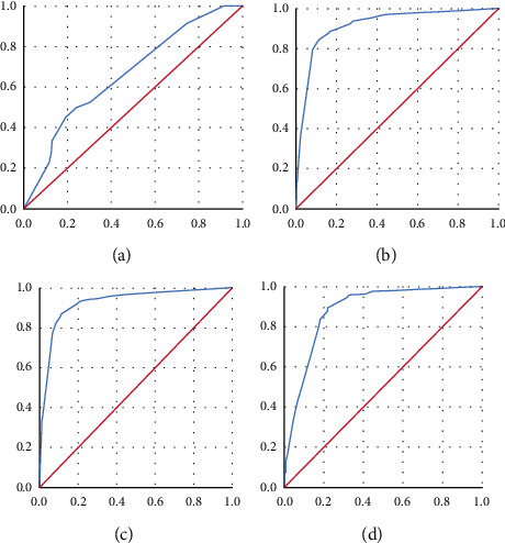
Results: The AUC-ROC score between CMP scores and B/W variables was 93,4%. The Spearman correlation between total deviation and pattern deviation was highly significant (p = 0,00). The correlation between AF GSS values and PRS was significant (p value = 0,00), the correlation between GSS of hyperautofluorescent points and PRS was significant (p value = 0,00), and the correlation between IR GSS and PRS was significant (p value = 0,00). The correlation between AF GSS and B/W variables was significant (p value = 0,002), the correlation between hyperautofluorescent points and B/W was not significant (p value = 0,40), and the correlation between IR GSS and B/W was significant (p = 0,00).
Conclusions: Based on our preliminary results, compass seems to be a reliable, quick, and safe device for the anatomical and functional study of GA. The direct visualization of the visual field on the fundus photography as a background allows a precise assessment and clinical monitoring of this disease.
Copyright © 2022 Roberta Farci et al.
Conflict of interest statement
The authors declare that they have no conflicts of interest.
Figures
References
LinkOut - more resources
Full Text Sources
Miscellaneous
