

Picosecond infrared laser driven sample delivery for simultaneous liquid-phase and gas-phase electron diffraction studies
- PMID: 36124204
- PMCID: PMC9482465
- DOI: 10.1063/4.0000159
Picosecond infrared laser driven sample delivery for simultaneous liquid-phase and gas-phase electron diffraction studies
Abstract
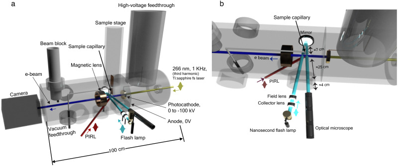
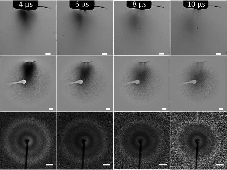
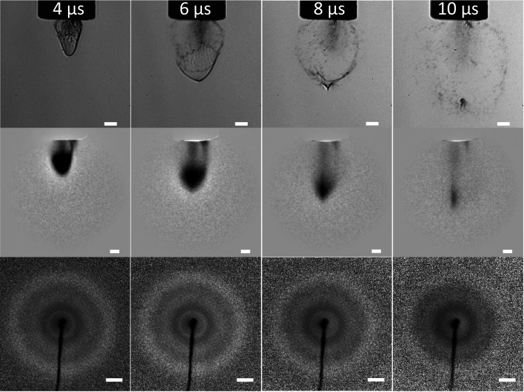
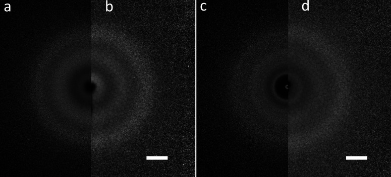
Here, we report on a new approach based on laser driven molecular beams that provides simultaneously nanoscale liquid droplets and gas-phase sample delivery for femtosecond electron diffraction studies. The method relies on Picosecond InfraRed Laser (PIRL) excitation of vibrational modes to strongly drive phase transitions under energy confinement by a mechanism referred to as Desorption by Impulsive Vibrational Excitation (DIVE). This approach is demonstrated using glycerol as the medium with selective excitation of the OH stretch region for energy deposition. The resulting plume was imaged with both an ultrafast electron gun and a pulsed bright-field optical microscope to characterize the sample source simultaneously under the same conditions with time synchronization equivalent to sub-micrometer spatial resolution in imaging the plume dynamics. The ablation front gives the expected isolated gas phase, whereas the trailing edge of the plume is found to consist of nanoscale liquid droplets to thin films depending on the excitation conditions. Thus, it is possible by adjusting the timing to go continuously from probing gas phase to solution phase dynamics in a single experiment with 100% hit rates and very low sample consumption (<100 nl per diffraction image). This approach will be particularly interesting for biomolecules that are susceptible to denaturation in turbulent flow, whereas PIRL-DIVE has been shown to inject molecules as large as proteins into the gas phase fully intact. This method opens the door as a general approach to atomically resolving solution phase chemistry as well as conformational dynamics of large molecular systems and allow separation of the solvent coordinate on the dynamics of interest.
© 2022 Author(s).
Figures
Similar articles
-
Molecular dynamics investigation of desorption and ion separation following picosecond infrared laser (PIRL) ablation of an ionic aqueous protein solution.J Chem Phys. 2016 Nov 28;145(20):204202. doi: 10.1063/1.4967164. J Chem Phys. 2016. PMID: 27908131
-
Homogenization of tissues via picosecond-infrared laser (PIRL) ablation: Giving a closer view on the in-vivo composition of protein species as compared to mechanical homogenization.J Proteomics. 2016 Feb 16;134:193-202. doi: 10.1016/j.jprot.2015.12.029. Epub 2016 Jan 8. J Proteomics. 2016. PMID: 26778141 Free PMC article.
-
Towards instantaneous cellular level bio diagnosis: laser extraction and imaging of biological entities with conserved integrity and activity.Nanotechnology. 2015 Jul 17;26(28):284001. doi: 10.1088/0957-4484/26/28/284001. Epub 2015 Jun 26. Nanotechnology. 2015. PMID: 26111866
-
How to make big molecules fly out of liquid water: applications, features and physics of laser assisted liquid phase dispersion mass spectrometry.Phys Chem Chem Phys. 2007 Jul 14;9(26):3335-60. doi: 10.1039/b615114k. Epub 2007 Mar 27. Phys Chem Chem Phys. 2007. PMID: 17664960 Review.
-
[Perspectives of laser-assisted keratoplasty: current overview and first preliminary results with the picosecond infrared laser (λ = 3 µm)].Ophthalmologe. 2014 Jun;111(6):523-30. doi: 10.1007/s00347-013-2995-7. Ophthalmologe. 2014. PMID: 24942118 Review. German.
References
-
- Yang J., Guehr M., Vecchione T., Robinson M. S., Li R., Hartmann N., Shen X., Coffee R., Corbett J., Fry A., Gaffney K., Gorkhover T., Hast C., Jobe K., Makasyuk I., Reid A., Robinson J., Vetter S., Wang F., Weathersby S., Yoneda C., Wang X., and Centurion M., “ Femtosecond gas phase electron diffraction with MeV electrons,” Faraday Discuss. 194, 563–581 (2016).10.1039/C6FD00071A - DOI - PubMed
LinkOut - more resources
Full Text Sources