Review
. 2022 Sep;3(3):031306.
doi: 10.1063/5.0096420. Epub 2022 Sep 13.

In vitro skeletal muscle models for type 2 diabetes

Affiliations
Review

In vitro skeletal muscle models for type 2 diabetes

Christina Y Sheng et al. Biophys Rev (Melville). 2022 Sep.

Abstract

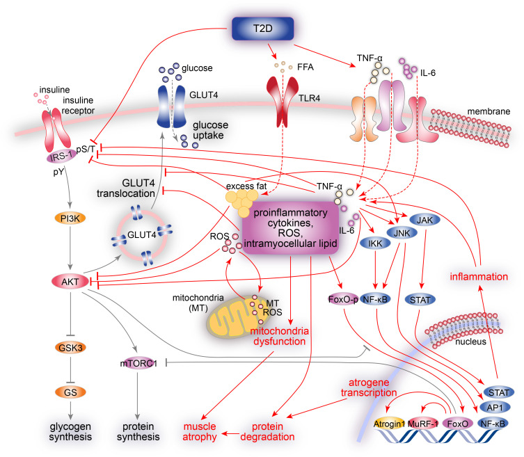
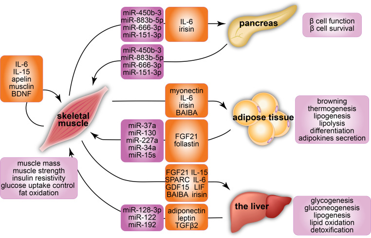
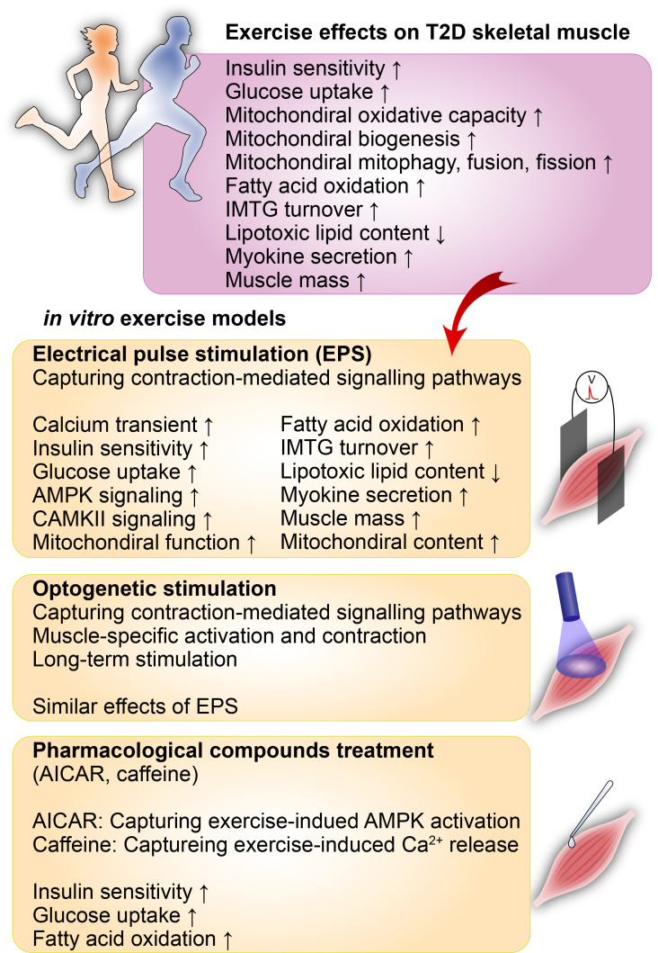
Type 2 diabetes mellitus, a metabolic disorder characterized by abnormally elevated blood sugar, poses a growing social, economic, and medical burden worldwide. The skeletal muscle is the largest metabolic organ responsible for glucose homeostasis in the body, and its inability to properly uptake sugar often precedes type 2 diabetes. Although exercise is known to have preventative and therapeutic effects on type 2 diabetes, the underlying mechanism of these beneficial effects is largely unknown. Animal studies have been conducted to better understand the pathophysiology of type 2 diabetes and the positive effects of exercise on type 2 diabetes. However, the complexity of in vivo systems and the inability of animal models to fully capture human type 2 diabetes genetics and pathophysiology are two major limitations in these animal studies. Fortunately, in vitro models capable of recapitulating human genetics and physiology provide promising avenues to overcome these obstacles. This review summarizes current in vitro type 2 diabetes models with focuses on the skeletal muscle, interorgan crosstalk, and exercise. We discuss diabetes, its pathophysiology, common in vitro type 2 diabetes skeletal muscle models, interorgan crosstalk type 2 diabetes models, exercise benefits on type 2 diabetes, and in vitro type 2 diabetes models with exercise.


Conflict of interest statement

The authors have no conflicts to disclose.

Figures

FIG. 1.
FIG. 1.
A summary of T2D pathophysiology in the skeletal muscle. In the healthy skeletal muscle (black arrows), insulin stimulates intracellular glucose metabolism by tyrosine phosphorylation (pY) of insulin receptors and IRSs. IRS proteins activate the PI3K/AKT pathway and promote many insulin actions via the activation of AKT by (1) increasing glucose influx via the translocation of GLUT4, (2) promoting protein synthesis via mTORC1 activation, (3) preventing the transcription of FoxO-dependent atrogene via FoxO phosphorylation, and (4) promoting glycogen synthesis via GSK3 inactivation. In the T2D skeletal muscle (red arrows), the inhibitory Ser/Thr phosphorylation (pS/T) of IRS-1 impairs its tyrosine phosphorylation (pY), resulting in the development of insulin resistance by impairing PI3K/AKT signaling, decreasing GLUT4 translocation and glucose transport, and impairing glucose phosphorylation and synthesis. In addition, altered phosphorylation of IRS-1 and reduced PI3K activity exacerbates muscle atrophy via the inhibition of mTORC1 signaling and activation of FoxO-dependent atrogene transcription (Atrogin1 and MuRF1). Increased ROSs, intramyocellular lipids, and proinflammatory cytokines (TNF-α and IL-6) lead to mitochondria dysfunction, lipotoxicity, and chronic inflammation via the activation of JNK, IKK/NF-κB, and JAK–STAT stress kinase pathways, resulting in Ser/Thr phosphorylation (pS/T) of insulin receptors and IRS proteins, further contributing to insulin resistance.
FIG. 2.
FIG. 2.
A summary of interorgan crosstalk via microRNAs and myokines with a focus on the skeletal muscle. The microRNAs and myokines are key factors secreted by the skeletal muscle that mediate the interaction between skeletal muscle and other organs, including the pancreas, adipose tissue, and the liver. The crosstalk of the skeletal muscle (1) enhances muscle mass and strength, insulin resistivity, glucose uptake control, and fat oxidation in skeletal muscles; (2) improves the function and survival of β-cells; (3) induces browning, thermogenesis, lipogenesis, lipolysis, differentiation, and adipokines secretion in adipose tissues; and (4) promotes glycogenesis, gluconeogenesis, lipogenesis, lipid oxidation, and detoxification in the liver.
FIG. 3.
FIG. 3.
In vitro exercise models for the study of T2D pathophysiology. Various in vitro exercise models have enabled mechanistic studies on numerous preventative and therapeutic effects of exercise on diabetes. Electrical pulse stimulation (EPS) of cultured myotubes has been commonly used to capture muscle-contraction-mediated signaling pathways. The optogenetic stimulation model provides an alternative method to activate contraction-mediated signaling pathways and enable prolonged stimulation and muscle maturation. Pharmacological compounds such as AICAR and AMPK have been used to study the effects of exercise on T2D in in vitro T2D models. AICAR has been used to mimic the exercise-induced activation of AMPK. In contrast, caffeine treatment has been applied to capture exercise-induced Ca2+ release from the sarcoplasmic reticulum and the downstream effects.

Similar articles

Cited by

References

    1. Galicia-Garcia U., Benito-Vicente A., Jebari S., Larrea-Sebal A., Siddiqi H., Uribe K. B., Ostolaza H., and Martin C., Int. J. Mol. Sci. 21(17), 6275 (2020).10.3390/ijms21176275 - DOI - PMC - PubMed
    1. Abdelalim E. M., Cell Mol. Life Sci. 78(6), 2459–2483 (2021).10.1007/s00018-020-03710-9 - DOI - PMC - PubMed
    1. Kahn S. E., Hull R. L., and Utzschneider K. M., Nature 444(7121), 840–846 (2006).10.1038/nature05482 - DOI - PubMed
    1. Abdul-Ghani M. A. and DeFronzo R. A., J. Biomed. Biotechnol. 2010, 476279.10.1155/2010/476279 - DOI - PMC - PubMed
    1. Whillier S., Adv. Exp. Med. Biol. 1228, 137–150 (2020).10.1007/978-981-15-1792-1 - DOI - PubMed

LinkOut - more resources