. 2022 Sep 23;50(17):9838-9857.
doi: 10.1093/nar/gkac766.

The high mobility group protein HMG20A cooperates with the histone reader PHF14 to modulate TGFβ and Hippo pathways

Affiliations

The high mobility group protein HMG20A cooperates with the histone reader PHF14 to modulate TGFβ and Hippo pathways

Elena Gómez-Marín et al. Nucleic Acids Res. .

Abstract

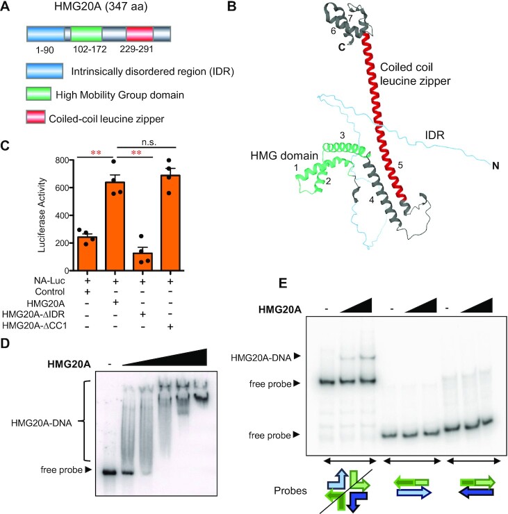
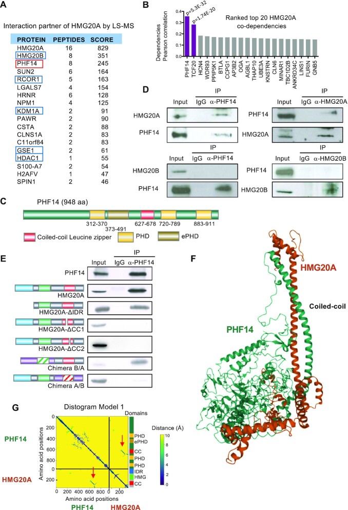
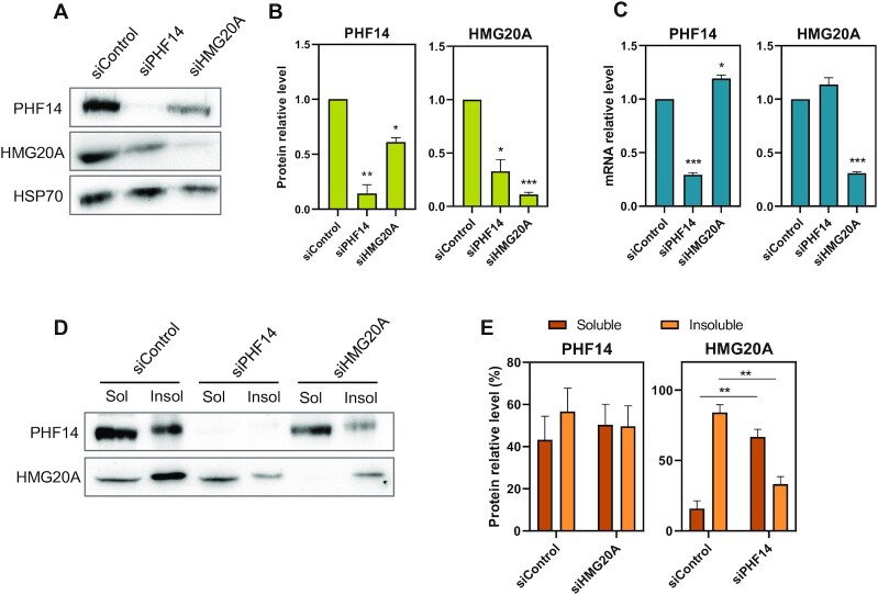
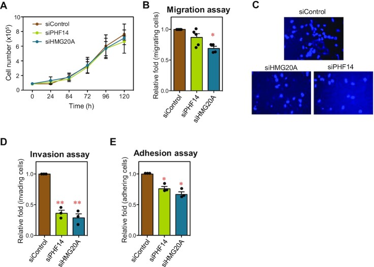
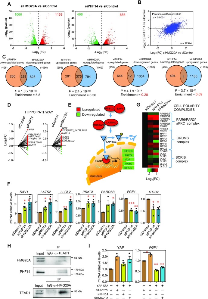
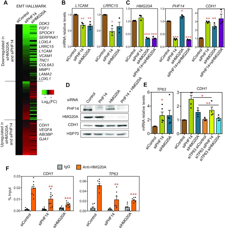
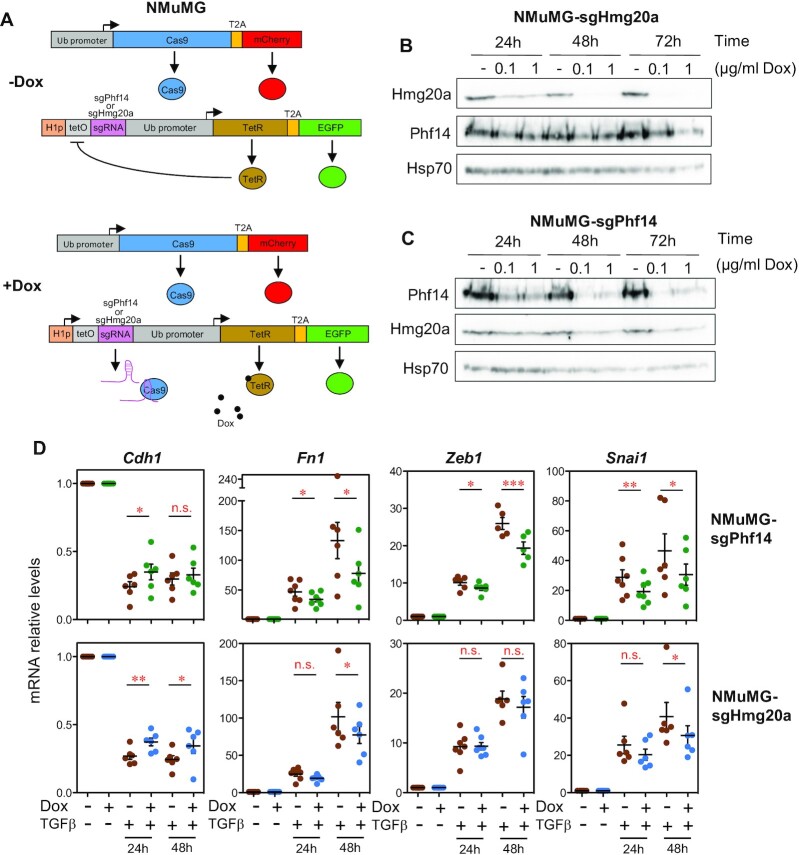
High mobility group (HMG) proteins are chromatin regulators with essential functions in development, cell differentiation and cell proliferation. The protein HMG20A is predicted by the AlphaFold2 software to contain three distinct structural elements, which we have functionally characterized: i) an amino-terminal, intrinsically disordered domain with transactivation activity; ii) an HMG box with higher binding affinity for double-stranded, four-way-junction DNA than for linear DNA; and iii) a long coiled-coil domain. Our proteomic study followed by a deletion analysis and structural modeling demonstrates that HMG20A forms a complex with the histone reader PHF14, via the establishment of a two-stranded alpha-helical coiled-coil structure. siRNA-mediated knockdown of either PHF14 or HMG20A in MDA-MB-231 cells causes similar defects in cell migration, invasion and homotypic cell-cell adhesion ability, but neither affects proliferation. Transcriptomic analyses demonstrate that PHF14 and HMG20A share a large subset of targets. We show that the PHF14-HMG20A complex modulates the Hippo pathway through a direct interaction with the TEAD1 transcription factor. PHF14 or HMG20A deficiency increases epithelial markers, including E-cadherin and the epithelial master regulator TP63 and impaired normal TGFβ-trigged epithelial-to-mesenchymal transition. Taken together, these data indicate that PHF14 and HMG20A cooperate in regulating several pathways involved in epithelial-mesenchymal plasticity.


Figures

Figure 1.
Figure 1.
Structural elements of the HMG20A protein. (A) Schematic depiction of the domain architecture of human HMG20A. (B) HMG20A structure as predicted by AlphaFold2. Structural elements depicted are named and represented with the same colors as in A. Alpha helices are numbered from 1 to 7. (C) Reporter assays showing activation of the SCNA2 promoter by HMG20A in HEK293T cells. Cells were transfected with empty vector (control) or expression vectors encoding the indicated proteins. Data are the average of four determinations from two independent experiments ± SE. Significance of the difference between distributions was determined using two-tailed Student’s t-tests. **P < 0.01. (D) Electrophoretic mobility shift assays (EMSA) of a double-stranded synthetic probe with increasing quantities (0.06, 0.3, 0.6, 1.5 and 3 μg) of purified His-HMG20A. (E) EMSA of four-way junction (4WJ) DNA structures with His-HMG20A (0.6 and 1.5 μg), in the presence of an excess of linear competitor DNA (poly(dI-dC)). As a control, EMSA was also performed with linear double-stranded probes containing the same sequence than the 4WJ structure. Schematics of the probes structure are shown.
Figure 2.
Figure 2.
HMG20A interacts with PHF14 through the coiled-coil domain. (A) Mass spectrometry analysis of proteins co-purified with HMG20A. Peptides from these proteins were exclusively identified in HMG20A-immunoprecipitation but not in mock control. Chaperones and common proteomic contaminants are excluded from the list. Subunits of the LSD1/CoREST complex are in blue boxes. (B) Top 20 co-dependencies of HMG20A. Depicted values are Pearson coefficients of the correlation between dependencies of HMG20A gene in 940 cancer cell lines, versus those of the indicated genes (data from Depmap Portal). Pearson coefficients with P value ≤ 10–10 are shown in purple. (C) Schematic showing the domain architecture of human PHF14. (D) Co-immunoprecipitation of endogenous HMG20A or HMG20B with PHF14. Cell extracts were subjected to immunoprecipitation with the indicated antibody or with IgG bulk antibodies as control. (E) PHF14 interacts with the coiled-coil domain of HMG20A. HEK293T cells were transfected with expression vectors encoding wild-type or the indicated mutant versions of HMG20A. Cell extracts were subjected to immunoprecipitation with anti-PHF14 antibody or with IgG bulk antibodies as control. (F) Alphafold2_multimers prediction of the PHF14-HMG20A complex through the formation of a two-stranded alpha-helical coiled-coil. (G) Distogram showing predicted relative distances between the amino acids of HMG20A and PHF14 for the structural model shown in F. A stretch of short distances corresponding to the contacts predicted in the two-stranded alpha-helical coiled-coil are visible (indicated with red arrows). A schematic showing the domain architecture of HMG20A and PHF14 is represented on the right of the plot.
Figure 3.
Figure 3.
Protein levels of HMG20A decreased in the absence of PHF14. (A) Determination by immunoblotting of PHF14 and HMG20A protein levels upon treatment of MDA-MB-231 cells with siPHF14, siHMG20A or a scrambled siRNA (siControl) for 48 h. HSP70 protein levels were also determined as a control. (B) Quantification of PHF14 and HMG20A protein levels from three independent immunoblotting experiments. Data were normalized with respect to HSP70. (C) Determination of the HMG20A and PHF14 mRNA by RT-qPCR in cells treated with siControl, siHMG20A or siPHF14. Data were normalized with respect to GAPDH mRNA levels. (D) MDA-MB-231 cells were treated with siPHF14, siHMG20A or siControl during 48 h. After fractionation, the presence of PHF14 and HMG20A proteins in the soluble (Sol) and insoluble (Insol) fractions was detected by immunoblotting. (E) Quantification of the PHF14 or HMG20A levels present in the soluble or insoluble fractions, as obtained in three independent experiment. (B, C, E) Data are the average of three independent experiments ± SE. Significance of the difference between distributions was determined using two-tailed Student’s t-tests. *P < 0.05; **P < 0.01, ***P < 0.001.
Figure 4.
Figure 4.
HMG20A and PHF14-depleted cells display common phenotypes. (A) Growth curve of MDA-MB-231 cells treated with siControl, siHMG20A or siPHF14. (B) Wound-healing assays were performed with MDA-MB-231 cells treated for 48 h with the indicated siRNAs. Quantification of the assays is represented. (C) Transwell invasion analyses of cells treated during 48 h with the indicated siRNAs. Micrographs of DAPI-stained cells migrated to the opposite side of the transwell membrane are shown. (D) Quantification of the invasion assays. (E) Quantification of the cell-cell adhesion assays under the indicated conditions. (A, B, D, E) Values are the average of three to five independent experiments ± SE. Significance of the differences with respect to the control were determined using two-tailed Student’s t-tests. *P ≤ 0.05; **P ≤ 0.01.
Figure 5.
Figure 5.
PHF14 or HMG20A depletion activates the Hippo pathway. MDA-MB-231 cells treated with siPHF14, siHMG20A or siControl for 48 h were analyzed by RNA-seq. (A) Volcano plots of the RNA-seq data at the indicated conditions. Significant deregulated genes (adjusted p value < 0.05 and |log2(FC)| > 0.5) are depicted in red (upregulated) or green (downregulated). Numbers inside the plots indicate downregulated or upregulated genes. (B) Correlation between gene expression changes (log2(FC)) after HMG20A or PHF14 KD. (C) Venn diagrams representing overlapping of deregulated genes after knocking down the indicated genes. Enrichment with respect to the randomly expected values, and probability of the result (P) assuming an hypergeometric distribution, are also indicated for the different comparisons. (D) Before–after plots showing changes of mRNA levels (log2(FC)) in Hippo pathway genes after KD of PHF14 (left panel) or HMG20A (right panel). (E) Schematic depiction of the Hippo pathway. Proteins in red or green are upregulated or downregulated, respectively, after HMG20A and PHF14 KD. (F) Determination of SAV1, LATS2, LLGL2, PRKCI, PARD6B, FGF1 and ITGB2 mRNA levels by RT-qPCR in MDA-MB-231 cells at 48 h after transfection with the indicated siRNAs. (G) Heatmap showing changes of mRNA levels (log2(FC)) of genes encoding proteins of cell polarity complexes. (H) Co-immunoprecipitation of endogenous HMG20A and PHF14 with TEAD1. Cell extracts were subjected to immunoprecipitation with the indicated antibody or with IgG bulk antibodies as control. (I) MDA-MB-231 were transfected with the indicated siRNAs and 24 h later infected with retrovirus expressing YAP-5SA protein. 48h later samples were processed for RNA isolation and YAP and FGF1 mRNA levels were determines by RT-qPCR. (F, I) Values are the average of three to five independent experiments ± SE. Significance of the differences with respect to the control were determined using two-tailed Student’s t-tests. *P ≤ 0.05; **P ≤ 0.01; ***P ≤ 0.001.
Figure 6.
Figure 6.
HMG20A KD and PHF14 KD MDA-MB-231 cells display decreased mesenchymal and increased epithelial markers. (A) Heatmap showing changes of mRNA levels (log2(FC)) in genes encoding EMT hallmark proteins. (B, C) Determination of L1CAM, and LRR15 mRNAs (B), or PHF14, HMG20A, and CDH1 mRNAs (C) by RT-qPCR in MDA-MB-231 cells 48 h after transfection with the indicated siRNAs. (D) Immunoblot analysis of PHF14, HMG20A and E-cadherin (CDH1) proteins in MDA-MB-231 cells treated during 48 h with the indicated siRNAs. HSP70 levels were also determined as control. (E) Determination of TP63 or CHD1 mRNAs by RT-qPCR in MDA-MB-231 cells at 48 h after transfection with the indicated siRNAs. (F) HMG20A binds to the CDH1 and TP63 promoters. MDA-MD-231 cells transfected with siControl, siHMG20A or siPHF14 were subjected to chromatin immunoprecipitation (ChIP) assays with anti-HMG20A and bulk IgG (control) antibodies. All data points of two (for TP63) or three (for CDH1) biological replicates with three technical replicate each are shown. (B, C, E) Values are the average of three to five independent experiments ± SE. (B, C, E, F) Significance of the differences with respect to the control were determined using two-tailed Student’s t-tests. *P ≤ 0.05; **P ≤ 0.01; ***P < 0.001.
Figure 7.
Figure 7.
Inducible CRISPR/Cas9 Phf14 or Hmg20a mutants in mouse NMuMG cells display impaired EMT. (A) Diagram depicting the Dox-inducible CRISPR/Cas9 system used to generate Phf14 and Hmg20a mutant NMuMG cell lines (28). (B, C) Immunoblotting showing Phf14 and Hmg20a protein levels, at the indicated times, after induction of CRISPR/Cas9-dependent mutagenesis of Phf14 and Hmg20a genes by addition of doxycycline (Dox). (D) Evolution of EMT markers upon induction of EMT by TGFβ addition to wild type (-Dox) or Phf14 and Hmg20a induced mutants (+Dox). Levels of Cdh1, Fn1, Zeb1 and Snai1 mRNA were determined by RT-qPCR at the indicated times. Values are the average of six to seven independent experiments ± SE. Significance of the differences with respect to the control were determined using two-tailed Student’s t-tests. *P ≤ 0.05; **P ≤ 0.01; ***P < 0.001.

Similar articles

Cited by

References

    1. Wynder C., Hakimi M.A., Epstein J.A., Shilatifard A., Shiekhattar R.. Recruitment of MLL by HMG-domain protein iBRAF promotes neural differentiation. Nat. Cell Biol. 2005; 7:1113–1117. - PubMed
    1. Hakimi M.A., Bochar D.A., Chenoweth J., Lane W.S., Mandel G., Shiekhattar R.. A core-BRAF35 complex containing histone deacetylase mediates repression of neuronal-specific genes. Proc. Natl. Acad. Sci. U.S.A. 2002; 99:7420–7425. - PMC - PubMed
    1. Lee M.G., Wynder C., Cooch N., Shiekhattar R.. An essential role for CoREST in nucleosomal histone 3 lysine 4 demethylation. Nature. 2005; 437:432–435. - PubMed
    1. Rivero S., Ceballos-Chavez M., Bhattacharya S.S., Reyes J.C.. HMG20A is required for SNAI1-mediated epithelial to mesenchymal transition. Oncogene. 2015; 34:5264–5276. - PubMed
    1. Ceballos-Chavez M., Rivero S., Garcia-Gutierrez P., Rodriguez-Paredes M., Garcia-Dominguez M., Bhattacharya S., Reyes J.C.. Control of neuronal differentiation by sumoylation of BRAF35, a subunit of the LSD1-CoREST histone demethylase complex. Proc. Natl. Acad. Sci. U.S.A. 2012; 109:8085–8090. - PMC - PubMed

Publication types