Review
. 2023 Feb;96(1142):20220207.
doi: 10.1259/bjr.20220207. Epub 2022 Sep 26.

Reporting and management of incidental lung findings on computed tomography: beyond lung nodules

Affiliations
Review

Reporting and management of incidental lung findings on computed tomography: beyond lung nodules

Carolyn Horst et al. Br J Radiol. 2023 Feb.

Abstract

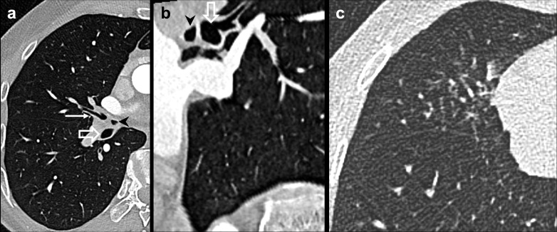
Non-nodular incidental lung findings can broadly be categorised as airway- or airspace-related abnormalities and diffuse parenchymal abnormalities. Airway-related abnormalities include bronchial dilatation and thickening, foci of low attenuation, emphysema, and congenital variants. Diffuse parenchymal abnormalities relate to the spectrum of diffuse parenchymal lung diseases cover a spectrum from interstitial lung abnormalities (ILAs) and pulmonary cysts to established diffuse parenchymal lung abnormalities such as the idiopathic interstitial pneumonias and cystic lung diseases. In this review, we discuss the main manifestations of these incidental findings, paying attention to their prevalence and importance, descriptors to use when reporting, the limits of what can be considered "normal", and conclude each section with some pragmatic reporting recommendations. We also highlight technical and patient factors which can lead to spurious abnormalities.


Figures

Figure 1.
Figure 1.
Table and diagram detailing literature search for this manuscript.
Figure 2.
Figure 2.
Bronchial calibre assessment. (a) Spuriously increased BAR in the anterior right upper lobe, as the subsegmental bronchus (arrow) is obliquely oriented relative to the homologous pulmonary arteriole, which has also just bifurcated (arrowheads). (b) A single subsegmental bronchus (arrow) which is slightly larger than the accompanying artery within 2 cm of the pleura can be ignored. (c) Established cylindrical bronchiectasis in a 79-year-old female with subsequently proven non-tuberculous mycobacterial infection. Note the tree-in-bud nodularity in the medial right lower lobe (block arrow). BAR, bronchoarterial ratio.
Figure 3.
Figure 3.
Single thickened subsegmental bronchus in the medial left lower lobe (arrow). There is at most trivial bronchial wall thickening and some atelectasis in the basal right lower lobe, both of which can probably be discounted.
Figure 4.
Figure 4.
Accessory cardiac bronchus in a 35-year-old male undergoing a CT coronary angiogram for unexplained dyspnoea and a family history of premature coronary artery disease. Coronary arteries were normal; however axial (a)and sagittal (b) 1 mm lung reconstructions demonstrate a blind-ending cardiac bronchus (black arrowhead) arising opposite the middle lobe bronchus (arrow), and anteromedial to the right lower lobe bronchus (block arrow). (c) Wide field of view 1 mm lung reconstructions show tree-in-bud nodularity in the middle lobe, indicating that in this case the cardiac bronchus may be acting as a sump for mucus accumulation and reaspiration in the context of the patient’s symptoms; however, the vast majority of patients with this variant are completely asymptomatic.
Figure 5.
Figure 5.
Examples of low-attenuation areas in three different individuals. (a) Solitary focus of low attenuation in the medial lingula (arrow), with relatively decreased pulmonary arterial calibre; this may be the result of localised small airways obstruction but is often encountered and best ignored. (b) 44-year-old obese female. 1 mm axial expiratory HRCT slice shows patches of decreased attenuation, but not exceeding the 25% upper limit of LAA which can be seen in normal patients. Note the apparently thickened subsegmental bronchi which is probably spurious due to the expiratory phase. (c) Upper and (d) lower axial HRCT slices from a 78-year-old female who is currently smoking, showing extensive mosaic attenuation—even on this inspiratory phase—with marked lobular pulmonary arterial calibre attenuation, representing extensive small airways obstruction in this context. With compatible obstructive pulmonary function, this would represent obliterative bronchiolitis. HRCT, high-resolution CT.
Figure 6.
Figure 6.
Pulmonary lucencies. (a) 8 mm poorly lucency has poorly defined walls. It is unclear if this is truly a “cyst” and may be a solitary focus of centrilobular emphysema, which can probably be ignored. (b) 7 mm slightly better-defined solitary pulmonary cyst in a 47-year-old male with no smoking history; such cysts are not uncommon over the age of 40 and can be ignored.
Figure 7.
Figure 7.
Different grades of CLE according to the Fleischner Society recommendations. (a) Trace CLE—scattered centrilobular lucencies (arrows) that overall occupy less than 0.5% of this upper lung zone. (b) Advanced destructive CLE with coalescent lucencies causing hyperexpansion and architectural distortion. CLE, centrilobular emphysema.
Figure 8.
Figure 8.
Paraseptal lucencies. (a) Approximately, four subpleural subcentimetre lucencies (circle) in the posteromedial right apex in a 55-year-old male with no smoking history. These can be ignored. (b) Moderate (>5% of the lung zone) centrilobular and substantial (mostly >1 cm subpleural blebs) paraseptal emphysema, along with some scarring in the lateral right upper lobe.
Figure 9.
Figure 9.
Spurious parenchymal appearances due to technical factors. (a) Unenhanced supine end-inspiratory HRCT 1 mm slice shows only dependent increased lung density. (b) However, on the CT pulmonary angiogram (CTPA) performed 2 min later, the presence of iodinated contrast, coupled with the gentle inspiration (or even expiration) phase that CTPA examinations are performed with, result in increased overall lung density and dependent high density. (c) In a different patient, unenhanced supine end-inspiratory HRCT 1 mm slice shows normal lung density, but a CTPA 9 min later (d) shows dependent atelectasis that was mistaken for pleural nodularity. CTPA, CT pulmonary angiogram
Figure 10.
Figure 10.
Incidentally detected fibrosing lung disease in a 78-year-old female. (a) Soft-tissue 1 mm CT slice on lung windows from the prone acquisition of a CT colonography examination demonstrates extensive ground-glass opacity as well as reticulation, with severe traction bronchiectasis. (b) The lung bases were normal on an abdominal CT years 8 years prior, indicating the disease had developed rapidly. A radiologic differential diagnosis of probable UIP or less likely mixed cellular/fibrotic NSIP was made; interestingly the patient also had joint stiffness, sicca symptoms and dysphagia, and a high antinuclear antibody titre of 1 in 320 but no other specific antibodies. Hence, the multidisciplinary team labelled this IPAFs, which is a designation for individuals with interstitial pneumonia and features suggestive of, but not definitive for, a defined connective tissue disease. IPAF, interstitial pneumonitis with autoimmune features; NSIP, non-specific interstitial pneumonia; UIP, usual interstitial pneumonia.
Figure 11.
Figure 11.
Diffuse pulmonary cysts. 54-year-old male with incidentally detected well-defined cysts in the upper zone (a), with a lower zone and slightly peripheral predominance (b). A diagnosis of Birt-Hogge-Dubé syndrome was suggested. (c) Subcentimetre cysts (circled) in a 45-year-old male who does not smoke. (d) Both the cysts and an area of peripheral decreased attenuation in the lateral left lower lobe are accentuated on a minimum intensity projection reconstruction (10 mm thickness, window width 500 HU, centre −856 HU). It transpired that the patient had a diagnosis of well-controlled asthma and normal lung function; the cysts were thus attributed to the recognised phenomenon of cysts associated with small airways disease.
Figure 12.
Figure 12.
Lung cancer associated with a cystic airspace in a 75-year-old female. (a) Sagittal reconstruction of a 3-mm-thick post-contrast CT chest demonstrates a thin walled multiseptated cyst in the right lower lobe (circle) (no higher-resolution reconstructions were performed). (b) Over 8 years, the cyst eventually developed a peripheral solid component and ground-glass nodularity, concerning for a lung cancer associated with a cystic airspace. Surgical resection proved a T1aN0M0 invasive pulmonary adenocarcinoma.
Figure 13.
Figure 13.
Defining ILAs in two different patients. Axial 1 mm HRCT slices from a 58-year-old male with no smoking history demonstrates barely perceptible ground-glass nodularity (arrowheads) in the upper zone (a), and minimal subpleural ground-glass opacity in the mid (b) and lower (c) zones (arrowheads in representative areas shown). The overall extent in any one zone is less than 5% of the lung area, so this would not qualify as an ILA. Axial 1 mm lung reconstructions from an arterially enhanced CT in a 60-year-old male who currently smokes demonstrates centrilobular emphysema with admixed peripheral ground-glass opacity, involving more than 5% of the the upper zone (d), with minimal subpleural ground-glass opacity in the mid (e) and lower (f) zones. This can be classified as a smoking-related ILA, but whether it represents subclinical vs early smoking-related interstitial lung disease depends on the respective absence or presence of symptoms and a physiologic deficit. (Images d, e and f courtesy of Dr Asia Ahmed, Consultant Thoracic Radiologist, University College London Hospital.)HRCT, high-resolution CT; ILA, interstitial lung abnormality.
Figure 14.
Figure 14.
Evaluation schema for incidentally detected pulmonary cysts, as recommended by the American College of Radiology. Reprinted from reference with kind permission of Elsevier. LAM, lymphangioleiomyomatosis; LIP, lymphoid interstitial pneumonia.

Similar articles

Cited by

References

    1. O’Sullivan JW, Muntinga T, Grigg S, Ioannidis JPA. Prevalence and outcomes of incidental imaging findings: umbrella review. BMJ 2018; 361: k2387. doi: 10.1136/bmj.k2387 - DOI - PMC - PubMed
    1. Vikgren J, Boijsen M, Andelid K, Ekberg-Jansson A, Larsson S, Bake B, et al. . High-resolution computed tomography in healthy smokers and never-smokers: a 6-year follow-up study of men born in 1933. Acta Radiol 2004; 45: 44–52. doi: 10.1080/02841850310002970 - DOI - PubMed
    1. Hatabu H, Hunninghake GM, Richeldi L, Brown KK, Wells AU, Remy-Jardin M, et al. . Interstitial lung abnormalities detected incidentally on CT: a position paper from the fleischner society. Lancet Respir Med 2020; 8: 726–37: S2213-2600(20)30168-5. doi: 10.1016/S2213-2600(20)30168-5 - DOI - PMC - PubMed
    1. Hansell DM. Thin-section CT of the lungs: the hinterland of normal. Radiology 2010; 256: 695–711. doi: 10.1148/radiol.10092307 - DOI - PubMed
    1. Aldington S, Shirtcliffe P, Nowitz M, Kingzett-Taylor A, Tweed M, Weatherall M, et al. . Incidental findings from lung CT scans: implications for research. J Med Imaging Radiat Oncol 2011; 55: 20–25. doi: 10.1111/j.1754-9485.2010.02224.x - DOI - PubMed