. 2022 Dec;43(12):2063-2078.
doi: 10.1002/humu.24471. Epub 2022 Oct 2.

Destabilization of mutated human PUS3 protein causes intellectual disability

Affiliations

Destabilization of mutated human PUS3 protein causes intellectual disability

Ting-Yu Lin et al. Hum Mutat. 2022 Dec.

Abstract

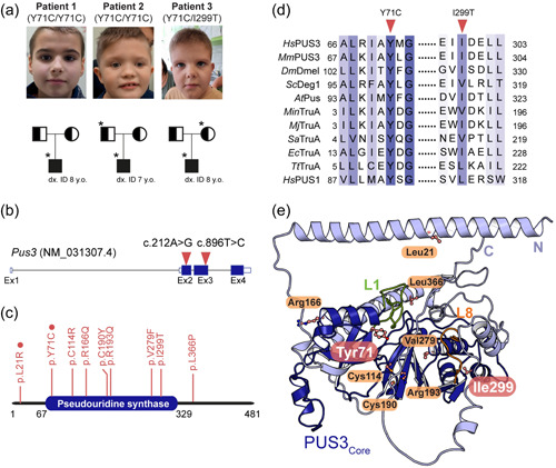
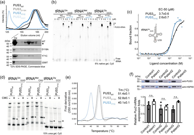
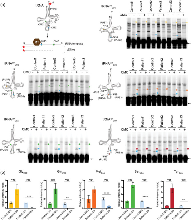
Pseudouridine (Ψ) is an RNA base modification ubiquitously found in many types of RNAs. In humans, the isomerization of uridine is catalyzed by different stand-alone pseudouridine synthases (PUS). Genomic mutations in the human pseudouridine synthase 3 gene (PUS3) have been identified in patients with neurodevelopmental disorders. However, the underlying molecular mechanisms that cause the disease phenotypes remain elusive. Here, we utilize exome sequencing to identify genomic variants that lead to a homozygous amino acid substitution (p.[(Tyr71Cys)];[(Tyr71Cys)]) in human PUS3 of two affected individuals and a compound heterozygous substitution (p.[(Tyr71Cys)];[(Ile299Thr)]) in a third patient. We obtain wild-type and mutated full-length human recombinant PUS3 proteins and characterize the enzymatic activity in vitro. Unexpectedly, we find that the p.Tyr71Cys substitution neither affect tRNA binding nor pseudouridylation activity in vitro, but strongly impair the thermostability profile of PUS3, while the p.Ile299Thr mutation causes protein aggregation. Concomitantly, we observe that the PUS3 protein levels as well as the level of PUS3-dependent Ψ levels are strongly reduced in fibroblasts derived from all three patients. In summary, our results directly illustrate the link between the identified PUS3 variants and reduced Ψ levels in the patient cells, providing a molecular explanation for the observed clinical phenotypes.

Keywords: PUS3; intellectual disorder; protein stability; pseudouridylation; tRNA modification.


Conflict of interest statement

The authors declare no conflict of interest.

Figures

Figure 1
Figure 1
Identification of the Y71C and I299T substitutions. (a) Pedigrees of the three individuals and their families. *The available samples for fibroblasts culture and further studies. The carriers are denoted as a symbol with half color whereas the patients are marked as a symbol filled with black. The age of genetic diagnosis of each patient is as indicated. (b) Schematic presentation of the human PUS3 transcript (red triangles indicate the point mutations). (c) Domain structure of the full‐length human PUS3 protein (a.a. 1‐481) where the pseudouridine synthase domain is as indicated (a.a. 67‐329). The reported causative missense variants are indicated, and the homozygotic mutations are highlighted with red circles. (d) Schematic presentation of the partial sequence alignment in human PUS3 and its homologs around the tyrosine 71 or the isoleucine 299 residues (indicated as triangles). TruA proteins include HsPUS3 (NP_112597.3), Drosophila melanogaster Dmel (NP_611646.1), Mus musculus Pus3 (NP_075781.3), Saccharomyces cerevisiae Deg1 (NP_116655.1), Methanocaldococcus infernus TruA (WP_013099720.1), Methanocaldococcus jannaschii TruA (WP_010871199.1), Staphylococcus aureus TruA (WP_075109215.1), EcTruA (QCJ59352.1), Arabidopsis thaliana Pus (NP_564438.1), and Thermus thermophilus TruA (WP_024118717.1). Human PUS1 sequence (NP_079491.2) was also aligned. (e) The cartoon representation of the predicted human PUS3 structure using Alphafold2 where the core domain is colored in deep blue and the N‐ and C‐termini are in light blue. The equivalent tRNA binding finger motifs (L1 and L8) of EcTruA are annotated as L1 (green, residue 71–88) and L8 (orange, residue 227–238) motifs in human PUS3. The predicted positions of all reported pathogenic PUS3 substitutions are indicated in ball and sticks presentation while the tyrosine 71 residue and isoleucine 299 residue are highlighted in pink. Oxygen is labeled in red while nitrogen is in blue.
Figure 2
Figure 2
The biochemical and biophysical characterizations of PUS3 proteins. (a) Gel filtration profiles of the recombinant HsPUS3, including PUS3WT, PUS3Y71C, PUS3I299T, and PUS3D118A, and the SDS‐PAGE gels of the corresponding eluted fractions of all proteins. *The protein identity is verified by mass spectrometry as a shorter version of PUS3. Inp: input. (b) The EMSA characterization of tRNA‐PUS3 complex formation. The unbound and bound tRNA are as annotated whereas the two different bound states are indicated (*, ** possible higher orders of PUS3‐tRNA complex). (c) Characterization of tRNA binding abilities of PUS3 by MST assays. The bound and unbound states of the fluorescent‐labeled tRNAGln UUG were plotted against the protein titrations (PUS3WT or PUS3Y71C). The measurements were triplicates and the average EC‐50 (μM) is listed in the inset. (d) Detection of PUS3‐dependent Ψ39 formation on various in vitro transcribed tRNAs. The reverse‐transcribed cDNA products were resolved in a 15% urea gel and the CMC‐Ψ stops the reverse transcription and results in shorter fragments which are indicated by *. Each tRNA primer (labeled with Cy5) is indicated by an arrow. (e) The melting temperatures (Tm) of PUS3 proteins. The intrinsic absorption at 350 and 330 nm were recorded, and the first derivative was calculated as the melting points of proteins and listed in the inset. (f) Western blot analysis of PUS3 protein levels and real‐time PCR analysis of PUS3 mRNA expression levels in the patients and healthy control‐derived fibroblasts. The HSP90 is the internal loading control for western blot while GUSB is the internal control for real‐time PCR. The relative amount of PUS3 mRNA was normalized to GUSB mRNA and is presented as fold change relative to control cells. Protein samples as well as mRNA samples were prepared from three independent cultures. Statistical analysis was performed using one‐way ANOVA (α = 0.05) with Sidak's multiple comparisons test. Data are presented as means ± SEM (n.s.: not significant). ANOVA, analysis of variance; MST, Microscale thermophoresis assay.
Figure 3
Figure 3
The Ψ profiles of various tRNAs in fibroblasts. (a) The scheme of detecting the presence of CMC‐Ψ using a reverse transcription method. The primer is labeled with Cy5 at its 5′ end. As the reverse transcriptase (RT) reverse transcribes the cDNA using the tRNA as a template, the presence of CMC‐Ψ causes RT to stop at the position before the CMC‐Ψ. As a result, various lengths of cDNA are generated according to the Ψ‐containing positions. The profile of Ψ sites based on the experimental validated MODOMICS database is presented in a cartoon presentation style (left to each gel). Each primer is base‐pairing at 3′ end of tRNA and it covers the Ψ55 position. Therefore, Ψ55 is undetectable in this assay and thus not included in the illustration. The reverse‐transcribed cDNA products were resolved in 15% urea gels and each PUS‐mediated CMC‐Ψ‐dependent fragments are indicated as * and colored coded (blue: Ψ39 by PUS3; green: Ψ26/27/28 by PUS1; yellow: Ψ13 by PUS7, orange: Ψ31 by PUS6, and red: Ψ35 by PUS1 or PUS7). Each tRNA primer is indicated by an arrow. (b) The triplicate measurements were performed. Statistical analysis was performed with two‐way ANOVA (α = 0.05) with Tukey's multiple comparisons test. Statistically significant differences are indicated (**p ≤ 0.01; ***p ≤ 0.001; ****p ≤ 0.0001). Data represent mean ± SEM. Ψ, pseudouridine; ANOVA, analysis of variance.

Similar articles

Cited by

References

    1. Abdelrahman, H. A. , Al‐Shamsi, A. M. , Ali, B. R. , & Al‐Gazali, L. (2018). A null variant in PUS3 confirms its involvement in intellectual disability and further delineates the associated neurodevelopmental disease. Clinical Genetics, 94, 586–587. 10.1111/cge.13443 - DOI - PubMed
    1. Adachi, H. , DeZoysa, M. D. , & Yu, Y. T. (2019). Detection and quantification of pseudouridine in RNA. Methods in Molecular Biology, 1870, 219–235. 10.1007/978-1-4939-8808-2_17 - DOI - PubMed
    1. Angelova, M. T. , Dimitrova, D. G. , Dinges, N. , Lence, T. , Worpenberg, L. , Carré, C. , & Roignant, J. Y. (2018). The emerging field of epitranscriptomics in neurodevelopmental and neuronal disorders. Frontiers in Bioengineering and Biotechnology, 6, 46. 10.3389/fbioe.2018.00046 - DOI - PMC - PubMed
    1. Begik, O. , Lucas, M. C. , Pryszcz, L. P. , Ramirez, J. M. , Medina, R. , Milenkovic, I. , Cruciani, S. , Liu H., Vieira H. G. S., Sas‐Chen A., Mattick J. S., Schwartz S., Novoa E. M. (2021). Quantitative profiling of pseudouridylation dynamics in native RNAs with nanopore sequencing. Nature Biotechnology, 39(10), 1278–1291. 10.1038/s41587-021-00915-6 - DOI - PubMed
    1. Boccaletto, P. , MacHnicka, M. A. , Purta, E. , Piątkowski, P. , Bagiński, B. , Wirecki, T. K. , De crécy‐Lagard, V. , Ross, R. , Limbach, P. A. , Kotter, A. , Helm, M. , & Bujnicki, J. M. (2018). MODOMICS: A database of RNA modification pathways. 2017 update. Nucleic Acids Research, 46(D1), D303–D307. 10.1093/nar/gkx1030 - DOI - PMC - PubMed

Publication types

MeSH terms