. 2022 Nov;121(11):3287-3303.
doi: 10.1007/s00436-022-07660-y. Epub 2022 Sep 20.

Role of cathepsin B of Naegleria fowleri during primary amebic meningoencephalitis

Affiliations

Role of cathepsin B of Naegleria fowleri during primary amebic meningoencephalitis

Itzel Berenice Rodríguez-Mera et al. Parasitol Res. 2022 Nov.

Abstract

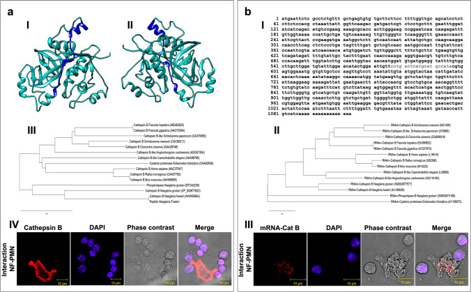
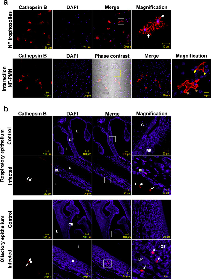
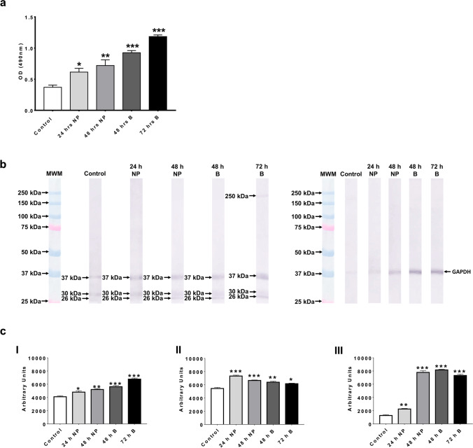
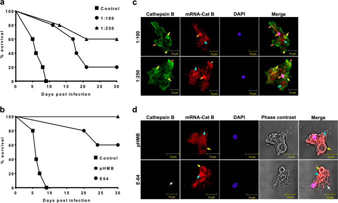
Naegleria fowleri causes primary amoebic meningoencephalitis in humans and experimental animals. It has been suggested that cysteine proteases of parasites play key roles in metabolism, nutrient uptake, host tissue invasion, and immune evasion. The aim of this work was to evaluate the presence, expression, and role of cathepsin B from N. fowleri in vitro and during PAM. Rabbit-specific polyclonal antibodies against cathepsin B were obtained from rabbit immunization with a synthetic peptide obtained by bioinformatic design. In addition, a probe was designed from mRNA for N. fowleri cathepsin B. Both protein and messenger were detected in fixed trophozoites, trophozoites interacted with polymorphonuclear and histological sections of infected mice. The main cathepsin B distribution was observed in cytoplasm or membrane mainly pseudopods and food-cups while messenger was in nucleus and cytoplasm. Surprisingly, both the messenger and enzyme were observed in extracellular medium. To determine cathepsin B release, we used trophozoites supernatant recovered from nasal passages or brain of infected mice. We observed the highest release in supernatant from recovered brain amoebae, and when we analyzed molecular weight of secreted proteins by immunoblot, we found 30 and 37 kDa bands which were highly immunogenic. Finally, role of cathepsin B during N. fowleri infection was determined; we preincubated trophozoites with E-64, pHMB or antibodies with which we obtained 60%, 100%, and 60% of survival, respectively, in infected mice. These results suggest that cathepsin B plays a role during pathogenesis caused by N. fowleri mainly in adhesion and contributes to nervous tissue damage.

Keywords: Naegleria fowleri; antibodies; cathepsin B; cysteine proteases; protease inhibitors.


Conflict of interest statement

The authors declare that they have no conflict of interest.

Figures

Fig. 1
Fig. 1
Specificity tests of N. fowleri cathepsin B. (a) SPCB-NF specificity. Ribbon representation of three-dimensional model of N. fowleri cathepsin B by in silico design. (I) front view, (II) back view. Selected peptide is shown in blue (amino acids 71–94). (III) Phylogenetic comparison of amino acid sequence of cathepsin B from various organisms. (IV) Immunodetection of SPCB-NF (red stain) in mouse bone marrow PMNs interacted with N. fowleri trophozoites, in blue the DNA staining. (b) Probe specificity. (I) N. fowleri cathepsin B mRNA nucleotide sequence. Nucleotide sequence selected for probe design is shown in red (cDNA position 577–596). (II) Phylogenetic comparison of nucleotide sequence of cathepsin B mRNA from various organisms. (III) Detection of cathepsin B mRNA probe (red stain) in mouse bone marrow PMNs interacted with N. fowleri trophozoites in blue the DNA staining. Trees was constructed by neighbor-joining method
Fig. 2
Fig. 2
Localization of cathepsin B in N. fowleri trophozoites (a) Immunodetection of cathepsin B in trophozoites alone or interacted with PMNs. Cathepsin B distribution in trophozoites alone was in cytoplasm (upper panel, white arrows), whereas trophozoite-PMNs interaction was in pseudopods and food-cups (lower panel, brown and yellow arrows, respectively). (b) Histological sections from uninfected and infected mice with N. fowleri trophozoites. Uninfected mice did not show cathepsin b staining in both the respiratory and olfactory epithelium (upper and lower panel control, respectively). On infected mice, trophozoites were in lumen before touching respiratory epithelial cells (upper panel infected, white arrows,) and migrating through olfactory epithelial cells (lower panel infected, white arrows). All of them with presence of cathepsin B. White boxes in merge represent area of magnification. Yellow boxes in phase contrast shown unstained trophozoites. L, lumen. C, cartilage. LP, lamina propria. RE, respiratory epithelium. OE, olfactory epithelium. Images are displayed at 40x (scale bar, 20 μm) and 100x (scale bar, 10 μm)
Fig. 3
Fig. 3
Expression of cathepsin B mRNA in N. fowleri trophozoites (a) mRNA expression in trophozoites alone or interacted with PMNs. mRNA expression in trophozoites alone was in nucleus and cytoplasm (upper panel, orange and turquoise arrow, respectively) and protein was in pseudopods and food-cups (upper panel, brown and yellow arrows, respectively), whereas mRNA in trophozoite-PMNs interaction was in cytoplasm (lower panel, turquoise arrow) and protein was in membrane (lower panel, brown arrow). Under this treatment, mRNA was outside the trophozoites in contact with PMNs alone (lower panel, pink arrows) or co-localized with protein (lower panel, gray arrows). (b) Histological sections from infected mice with N. fowleri trophozoites. Trophozoites were in lumen of nasal cavity of respiratory epithelium with mRNA expression and presence of cathepsin B (white and turquoise arrows, respectively). White boxes in merge represent area of magnification. Yellow boxes in phase contrast shown unstained trophozoites. L, lumen. RE, respiratory epithelium. Images are displayed at 40x (scale bar, 20 μm) and 100x (scale bar, 10 μm)
Fig. 4
Fig. 4
Release of mRNA and protein into extracellular medium by N. fowleri trophozoites. (a) mRNA release by trophozoites interacted with PMNs was localized in contact with PMNs alone or co-localized with cathepsin B (turquoise and gray arrows, respectively), whereas protein was in pseudopods a food-cups (brown and yellow arrows, respectively). (b) In tissue sections of infected mice, trophozoites expressed mRNA within small vesicles distributed in cytoplasm (blue arrows), but their release could not be observed, whereas cathepsin B release was observed within small vesicles (red arrows). (c) mRNA and protein distribution shown in 3D images (blue and white arrows, respectively). All 3D images were taken from the magnification of figure b. L, lumen. C, cartilage. RE, respiratory epithelium. Images are displayed at 40x (scale bar, 20 μm) and 100x (scale bar, 10 μm)
Fig. 5
Fig. 5
Release of cathepsin B from N. fowleri during PAM. (a) Culture supernatants of amoebae recovered from nasal passages (NP) or brain (B) were evaluated for cathepsin B release. All treatments showed a significant increase compared to control of which culture supernatants of B recovered trophozoites showed the highest release. (b) Molecular weight analysis of secreted proteins. 26, 30 and 37 kDa were the most immunogenic bands recognized in all treatments. GAPDH was used as a loading control. (c) Densitometric analysis of immunogenic bands evaluated with Image J software. (I) 37 kDa. (II) 30 kDa. (III) 26 kDa. All molecular weights evaluated by densitometry showed higher recognition in all treatments compared to the control. Significant differences versus control are indicated as follows: *p < .05, **p < .01 or ***p < .001
Fig. 6
Fig. 6
Participation of N. fowleri cathepsin B during PAM. (a) Trophozoites preincubated with anti-SPCB-NF antibodies provided 20 and 60% survival (1:100 and 1:250 dilutions, respectively). (b) Trophozoites preincubated with pHMB provide 100% survival, whereas E-64, 60%. (c) Integrity of trophozoites pre-incubated with antibodies. In both antibody dilutions, cathepsin B distribution could be observed in pseudopods and food-cups (brown and yellow arrows, respectively), whereas mRNA expression was in cytoplasm, nuclei, and pseudopods (turquoise, orange and brown arrows, respectively). (d) Integrity of trophozoites pre-incubated with pHMB or E-64. Cathepsin B could not be located under any treatment, but messenger was observed in food-cups and cytoplasm (yellow and turquois arrows, respectively). Images are displayed at 100x (scale bar, 10 μm)

References

    1. Aldape K, Huizinga H, Bouvier J, McKerrow J. Naegleria fowleri: characterization of a secreted histolytic cysteine protease. Exp Parasitol. 1994;78(2):230–241. doi: 10.1006/expr.1994.1023. - DOI - PubMed
    1. Allena R. Cell migration with multiple pseudopodia: temporal and spatial sensing models. Bull Math Biol. 2013;75(2):288–316. doi: 10.1007/s11538-012-9806-1. - DOI - PubMed
    1. Aurongzeb M, et al. In silico analysis of Naegleria fowleri cathepsin B paralogs: important drug targets. Eur Rev Med Pharmacol sci. 2021;25(8):3162–3172. doi: 10.26355/eurrev_202104_25725. - DOI - PubMed
    1. Barrett AJ, Brown MA. Chicken liver Pz-peptidase, a thiol-dependent metallo-endopeptidase. Biochem J. 1990;271(3):701–706. doi: 10.1042/bj2710701. - DOI - PMC - PubMed
    1. Betanzos A, Bañuelos C, Orozco E (2019) Host Invasion by Pathogenic Amoebae: Epithelial Disruption by Parasite Proteins. Genes 10(8). 10.3390/genes10080618 - PMC - PubMed

LinkOut - more resources