. 2022 Sep 20;18(9):e1010846.
doi: 10.1371/journal.ppat.1010846. eCollection 2022 Sep.

A member of the tryptophan-rich protein family is required for efficient sequestration of Plasmodium berghei schizonts

Affiliations

A member of the tryptophan-rich protein family is required for efficient sequestration of Plasmodium berghei schizonts

Julie-Anne Gabelich et al. PLoS Pathog. .

Abstract

Protein export and host membrane remodeling are crucial for multiple Plasmodium species to establish a niche in infected hosts. To better understand the contribution of these processes to successful parasite infection in vivo, we sought to find and characterize protein components of the intraerythrocytic Plasmodium berghei-induced membrane structures (IBIS) that form in the cytoplasm of infected erythrocytes. We identified proteins that immunoprecipitate with IBIS1, a signature member of the IBIS in P. berghei-infected erythrocytes. In parallel, we also report our data describing proteins that co-precipitate with the PTEX (Plasmodium translocon of exported proteins) component EXP2. To validate our findings, we examined the location of three candidate IBIS1-interactors that are conserved across multiple Plasmodium species, and we found they localized to IBIS in infected red blood cells and two further colocalized with IBIS1 in the liver-stage parasitophorous vacuole membrane. Successful gene deletion revealed that these two tryptophan-rich domain-containing proteins, termed here IPIS2 and IPIS3 (for intraerythrocytic Plasmodium-induced membrane structures), are required for efficient blood-stage growth. Erythrocytes infected with IPIS2-deficient schizonts in particular fail to bind CD36 as efficiently as wild-type P. berghei-infected cells and therefore fail to effectively sequester out of the circulating blood. Our findings support the idea that intra-erythrocytic membrane compartments are required across species for alterations of the host erythrocyte that facilitate interactions of infected cells with host tissues.


Conflict of interest statement

The authors have declared that no competing interests exist.

Figures
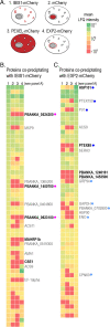
Fig 1
Fig 1. Immunoprecipitation of P. berghei proteins from infected red blood cells identifies proteins that co-precipitate specifically with IBIS1-mCherry and EXP2-mCherry.
A) The indicated mCherry-tagged proteins were immunoprecipitated from lysates of late stage P. berghei using RFP-specific nanobodies and co-precipitating proteins were identified by LC-MS/MS. B) P. berghei proteins that were detected in the IBIS1-mCherry sample in at least two of the experimental replicates are depicted in the heatmap, which indicate the mean normalized LFQ intensities of detected proteins of three independent experiments. Each row represents a protein detected in the IBIS1-mCherry eluates. The columns represent the immunoprecipitates prepared from the mCherry-expressing parasite lines indicated in (A) (1: IBIS1-mCherry, 2: mCherry, 3: PEXEL-mCherry, 4:EXP2-mCherry). Proteins that were enriched in the IBIS1-mCherry pulldown relative to at least two of the three controls (top 10% of the normal distribution) are annotated. Those in bold were enriched relative to all three controls. Proteins marked with a pink dot were followed up further in this study. C) P. berghei proteins that were detected in the EXP2-mCherry sample in at least two of the experimental replicates are depicted in the heatmap, which indicate the mean normalized LFQ intensities of detected proteins of three independent experiments. Each row represents a protein detected in the EXP2-mCherry eluates. The columns represent the immunoprecipitates prepared from the different mCherry-expressing parasite lines indicated in (A). Proteins marked with dark blue dots are confirmed to play roles in PTEX-mediated export, and those marked with light blue dots were previously found biochemically associated with components of the PTEX complex [33,34].
Fig 2
Fig 2. mCherry tagged PBANKA_0524300 (IPIS2), PBANKA_0623100 (IPIS3) colocalize with IBIS1-GFP at the liver-stage parasitophorous vacuole.
A) Protein features of candidate proteins that coprecipitated with IBIS1. B) Hepatoma cells were infected with sporozoites after the cross-fertilization of IBIS1-GFP and either PBANKA_0524300-mCherry, PBANKA_0623100-mCherry or PBANKA_1400700-mCherry transgenic parasite lines. Cells were fixed at indicated time points and the mCherry and GFP signals were amplified by using anti-RFP and anti-GFP antibodies. Scale bars, 10 μm.
Fig 3
Fig 3. PBANKA_0524300 (IPIS2), PBANKA_0623100 (IPIS3), PBANKA_1400700 and putative P. vivax orthologs localize to discrete structures in the cytoplasm of infected erythrocytes.
A) PBANKA_0524300-mCherry, PBANKA_0623100-mCherry, and PBANKA_1400700-mCherry colocalize with the IBIS marker IBIS1-GFP in the erythrocyte cytoplasm. Infected cells were fixed and labeled with anti-GFP and anti-mCherry antibodies to amplify the fluorescent signal. Scale bars, 5 μm. B) Erythrocytes infected with P. berghei expressing the HA-tagged P. vivax proteins PvTRAg8 or PvTRAg2, which are putative orthologs of IPIS2 and IPIS3, respectively, were fixed and labeled with anti-HA antibodies. Scale bars, 5 μm.
Fig 4
Fig 4. ipis2- and ipis3- contribute to blood-stage growth efficiency during P. berghei infection.
A) The appearance of ipis3- parasites in the blood following sporozoite injection is delayed in comparison to wild-type. C57BL/6 mice were infected with 1,000 sporozoites of either ipis2-, ipis3- or wild-type parasites, and blood-stage infections were monitored starting from day 3 post-infection with Giemsa-stained blood smears. Statistics: Log-rank (Mantel-cox) test; n = 5 (ipis2-), n = 10 (ipis3-). B) The growth of ipis2- and ipis3- parasites in the liver does not differ significantly from wild-type P. berghei. The relative parasite burden in the liver was measured by qPCR of RNA extracted from livers forty-two hours after intravenous injection of either ipis2-, ipis3-, or wild-type sporozoites. Levels of Pb18s rRNA were normalized to levels of mouse GAPDH RNA in each infected liver. Statistics: unpaired two-tailed t-test; n = 5; ns: not significant. C) The ipis2- and ipis3- lines do not grow as efficiently as wild type in the blood of co-infected animals. Mice were infected with equal amounts of wild-type P. berghei-infected erythrocytes and ipis2-[mCherry; PyrS] or ipis3-[mCherry; PyrS] infected erythrocytes. Parasitemia was measured by flow cytometry between days 4–7 after infection. * p <0.05, ** p < 0.01, *** p < 0.005 determined by unpaired two-tailed t-test; n = 5. D) The absence of one IPIS protein does not dramatically influence the expression level of the other. Erythrocyte-associated mCherry signal was quantified in fixed unstained infected cells. The mCherry signal intensity was plotted as a ratio to parasite size to account for differences in expression in different stages. In the ipis2- line, a statistically significant increase in mCherry signal was detected; however the extent was negligible. * p <0.05, unpaired two-tailed t-test.
Fig 5
Fig 5. IPIS2 contributes to efficient sequestration of schizonts from the peripheral circulation.
A) ipis2- or ipis3- infection increases spleen weight more than wild-type P. berghei infection. Mice were infected with sporozoites of either wild type (n = 10), ipis2-[GFP-Luc; PyrS] (n = 4), or ipis3-[GFP-Luc; PyrS] (n = 10) lines, and 8 days after infection the spleens were dissected and weighed. The spleen weights were plotted as ratios to mouse body weight.* p <0.05, unpaired two-tailed t-test. B) The absence of IPIS2 or IPIS3 resulted in increased schizonts in peripheral blood compared to wild-type P. berghei during synchronous infections. Mice were infected with P. berghei schizonts (wild-type, ipis2-[GFP-Luc; PyrS] or ipis3-[GFP-Luc; PyrS]) and the parasite stages detected in peripheral blood smears were quantified at the indicated time points following infection. n = 5 mice in three experimental replicates. Statistical annotations (* p <0.05, ** p < 0.01, *** p < 0.005) refer to percent of schizonts in the transgenic line in comparison to the wild-type line at the indicated time points, unpaired two-tailed t- test. C) Tissue-associated parasite material was assessed by luminescence in homogenized tissues 23 hours after infection with P. berghei schizonts (wild-type Pb GFP-Luc, ipis2-[GFP-Luc; PyrS] or ipis3-[GFP-Luc; PyrS]). Values are normalized to organs from wild-type-infected mice.** p < 0.01, **** p < 0.0001, ns: not significant, unpaired two-tailed t-test. D) Red blood cells infected with IPIS2-deficient P. berghei do not bind efficiently to CD36-expressing CHO cells. Schizonts were incubated with CHO cells and the number of infected-red blood cells bound per cell was quantified. The background binding to control CHO cells was subtracted, and the values were normalized to wild-type-infected red blood cell binding. The depicted data comprises three experimental replicates with three technical replicates for each parasite line and CHO cell line. ** p < 0.01, ns: not significant, unpaired two-tailed t-test.

Similar articles

Cited by

References

    1. de Koning-Ward TF, Gilson PR, Boddey JA, Rug M, Smith BJ, Papenfuss AT, et al.. A newly discovered protein export machine in malaria parasites. Nature. 2009; 459(7249):945–9. doi: 10.1038/nature08104 . - DOI - PMC - PubMed
    1. Beck JR, Muralidharan V, Oksman A, Goldberg DE. PTEX component HSP101 mediates export of diverse malaria effectors into host erythrocytes. Nature. 2014; 511(7511):592–5. doi: 10.1038/nature13574 . - DOI - PMC - PubMed
    1. Elsworth B, Matthews K, Nie CQ, Kalanon M, Charnaud SC, Sanders PR, et al.. PTEX is an essential nexus for protein export in malaria parasites. Nature. 2014; 511(7511):587–91. doi: 10.1038/nature13555 . - DOI - PubMed
    1. Coatney GR. The simian malarias: zoonoses, anthroponoses, or both? Am J Trop Med Hyg. 1971; 20(6):795–803. doi: 10.4269/ajtmh.1971.20.795 . - DOI - PubMed
    1. Aikawa M. Parasitological review. Plasmodium: the fine structure of malarial parasites. Exp Parasitol. 1971; 30(2):284–320. doi: 10.1016/0014-4894(71)90094-4 . - DOI - PubMed

Publication types