. 2022 Oct:84:104266.
doi: 10.1016/j.ebiom.2022.104266. Epub 2022 Sep 17.

Different oxytocin and corticotropin-releasing hormone system changes in bipolar disorder and major depressive disorder patients

Affiliations

Different oxytocin and corticotropin-releasing hormone system changes in bipolar disorder and major depressive disorder patients

Lei Guo et al. EBioMedicine. 2022 Oct.

Abstract

Background: Oxytocin (OXT) and corticotropin-releasing hormone (CRH) are both produced in hypothalamic paraventricular nucleus (PVN). Central CRH may cause depression-like symptoms, while peripheral higher OXT plasma levels were proposed to be a trait marker for bipolar disorder (BD). We aimed to investigate differential OXT and CRH expression in the PVN and their receptors in prefrontal cortex of major depressive disorder (MDD) and BD patients. In addition, we investigated mood-related changes by stimulating PVN-OXT in mice.

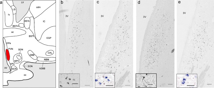
Methods: Quantitative immunocytochemistry and in situ hybridization were performed in the PVN for OXT and CRH on 6 BD and 6 BD-controls, 9 MDD and 9 MDD-controls. mRNA expressions of their receptors (OXTR, CRHR1 and CRHR2) were determined in anterior cingulate cortex and dorsolateral prefrontal cortex (DLPFC) of 30 BD and 34 BD-controls, and 24 MDD and 12 MDD-controls. PVN of 41 OXT-cre mice was short- or long-term activated by chemogenetics, and mood-related behavior was compared with 26 controls.

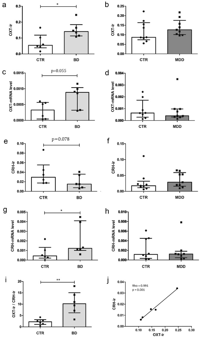
Findings: Significantly increased OXT-immunoreactivity (ir), OXT-mRNA in PVN and increased OXTR-mRNA in DLPFC, together with increased ratios of OXT-ir/CRH-ir and OXTR-mRNA/CRHR-mRNA were observed in BD, at least in male BD patients, but not in MDD patients. PVN-OXT stimulation induced depression-like behaviors in male mice, and mixed depression/mania-like behaviors in female mice in a time-dependent way.

Interpretation: Increased PVN-OXT and DLPFC-OXTR expression are characteristic for BD, at least for male BD patients. Stimulation of PVN-OXT neurons induced mood changes in mice, in a pattern different from BD.

Funding: National Natural Science Foundation of China (81971268, 82101592).

Keywords: Bipolar disorder; Corticotropin-releasing hormone; Hypothalamus; Major depressive disorder; Oxytocin; Prefrontal cortex.


Conflict of interest statement

Declaration of interests We declare no competing interests.

Figures

Figure 1
Figure 1
Immunocytochemistry and in situ hybridization images of oxytocin and corticotropin-releasing hormone in the human hypothalamic paraventricular nucleus. (a) Schematic representation of the nuclei of the human hypothalamus. Abbreviations: OX, optic chiasma, NBM: nucleus basalis of Meynert, hDBB: horizontal limb of the diagonal band of Broca, SDN: sexually dimorphic nucleus of the preoptic area, SCN: suprachiasmatic nucleus, BST: bed nucleus of the stria terminalis, (c = centralis; m = medialis; l = lateralis; p = posterior); PVN: paraventricular nucleus, SON: supraoptic nucleus; Dpe: periventricular nucleus dorsal zone, VPe: periventricular nucleus ventral zone, fx: fornix, 3V: third ventricle, ac: anterior commissure (From Swaab, Dick. (2003). The Human Hypothalamus. Basic and Clinical Aspects. Mycological Research. 79), please note that PVN is marked in red. (b-c) Overview and higher magnification images of oxytocin (OXT)-immunoreactivity (ir) (b) or OXT-mRNA (c) signal in the PVN. (d-e) Overview and higher magnification images of corticotropin-releasing hormone (CRH)-ir (d) or CRH-mRNA (e) signal in the PVN. Scale bar=200μm in the overviews of b-e; and scale bar=50μm in the higher magnification images of b-e.
Figure 2
Figure 2
Analysis on the expression of oxytocin and corticotropin-releasing hormone in the hypothalamic paraventricular nucleus in mood disorder patients and their controls. (a-b) Oxytocin (OXT)-ir showed a significant increase in bipolar disorder (BD) patients compared with their controls (p=0·037, Mann-Whitney U test, a), while no significant difference was present between major depressive disorder (MDD) and their controls (b). (c-d) A trend of increased OXT-mRNA (p=0·055, Mann-Whitney U test, c) was found in BD patients compared with their controls, while no significant difference in OXT-mRNA (d) was found between MDD patients and their controls. (e-f) PVN CRH-ir showed a trend of lower expression in BD patients compared with their controls (p=0·078, Mann-Whitney U test, e), but no significant difference between MDD patients and their controls (f). (g-h) Significantly increased CRH-mRNA (p=0·037, Mann-Whitney U test, g) was found in BD patients compared with their controls, but no significant difference in CRH-mRNA (h) was found between MDD patients and their controls. (i) The ratio of OXT-ir/CRH-ir was significantly higher in BD patients compared with their controls (p=0·006, Mann-Whitney U test). (j) Significant positive correlation was found between OXT-ir and CRH-ir in male BD patients (p=0·001, Spearman test). Columns are shown as median with interquartile range. CTR: control. * p <0·05.
Figure 3
Figure 3
Analysis on the mRNA expression of oxytocin receptor and corticotropin-releasing hormone receptors in the prefrontal cortex of mood disorder patients and their controls. (a-b) oxytocin receptor (OXTR)-mRNA levels in the dorsolateral prefrontal cortex (DLPFC) in bipolar disorder (BD) patients showed no significant difference compared with their controls (a), while male BD patients showed significantly increased OXTR-mRNA in DLPFC compared with male controls (p<0·01, Mann-Whitney U test) (b). (c-d) No significant change was found within OXTR-mRNA levels in DLPFC of major depressive disorder (MDD) patients and their controls. Still, no significant difference was found after male patients were separated from female patients. (e-f) No significant change of CRHR2-mRNA was found in the DLPFC in BD patients compared with their controls, and no sex difference was found. (g-h) A trend (p=0·056, Mann-Whitney U test) of lower CRHR2-mRNA was found in the DLPFC in MDD patients compared with their controls, and no sex difference was found. (i) Significantly higher ratio of OXTR-/CRHR1-mRNA was found in the DLPFC of male BD patients compared with male controls. (j-k) Significantly higher ratio of OXTR-mRNA/CRHR2-mRNA was found in both the DLPFC (j) and anterior cingulate cortex (ACC, k) of male BD patients compared with male controls. Columns are shown as median with interquartile range. ** p<0·01; * 0·01< p <0·05.
Figure 4
Figure 4
Significant mood-related behavior changes in mice with short-term oxytocin chemogenetic stimulation in hypothalamic paraventricular nucleus. (a-b) Virus (AAV-hSyn-DIO-hM3Dq-eGFP, activated group) or AAV-hSyn-DIO-eGFP (control group), were injected with the angle of 10° into hypothalamic paraventricular nucleus (PVN) (A-P: -1.0 mm, M-L: ±1.2mm, D-V: -5.1mm) at the speed of 60nl/min, 250nl/side. (c-e) Co-localization (yellow) of the virus signal (green) and c-fos-ir signal (red) in the control group (d) and chemogenetic activated group (e). Quantification analysis (c) showed that there was less co-localization in the control group than in the activated group. (f) Co-localization (yellow) of the AAV-DIO-hM3DGq virus signal (green) and oxytocin (OXT)-ir signal (red). The virus signal is fully co-existed with PVN-OXT signal as it shows in the highlight box, which indicates the virus expresses specifically in the OXT neurons and the injection place is correct. (g-h) Time schedule of short-term or long-term PVN-OXT stimulation and behavior tests. In the short term PVN-OXT stimulation group, behavior tests were conducted 15min after the first injection of clozapine N-oxide (CNO, i.p. g), while in the long term PVN-OXT stimulation schedule, behavior tests were conducted after at least 3 days of CNO injection (once a day, h), and behavior tests began at 15min after the last injection. (i-j) Significant behaviors changes observed in short term PVN-OXT stimulation. Male PVN-OXT-activated mice spent significantly less time in the central area in the open field test (OFT, p=0·039, Student t test) compared with male controls. The moving tracks of the median-level mouse of each group were presented (i). In the social interaction (SI) test, female PVN-OXT-activated mice showed more entries into the mouse chamber (p<0·01, Student t test) and more contact with the other mouse (p=0·018, Student t test), more entries into the object chamber (p=0·019, Student t test), and more total entries through three chambers (p<0·01, Student t test) compared with their female control mice. The heatmap showed the median level of the mouse movement in each group (j). Columns are shown as mean with SD. Scale bar=50μm in image d-f. ** p<0·01; * 0·01< p <0·05.
Figure 5
Figure 5
Significant mood-related behavior changes in mice with long-term oxytocin chemogenetic stimulation in hypothalamic paraventricular nucleus. (a) Male paraventricular nucleus (PVN)-oxytocin (OXT)-activated mice showed significantly less moving distance (p=0·028, Student t test) in the light zone in the light-dark box (LDB) compared with male control mice. The moving tracks of the median-level mouse of each group were presented. (b) In the social interaction (SI) test, male PVN-OXT-activated mice showed more time in the object chamber, more interactions with the object and more time interacting with the object (p≤0·021, Student t test) compared with male control mice. The heatmap showed movements of the mouse that was at the median level in each group. (c) Female PVN-OXT-activated mice showed significantly more entries (p<0·01, Student t test) to the central area in the open field test (OFT) compared with female control mice. The moving tracks of the median-level mouse of each group were presented. (d) Female PVN-OXT-activated mice showed significantly more immobile time (p=0·025, Student t test) in the forced swim test (FST). (e) Female PVN-OXT-activated mice showed less time interacting with the mouse (p=0·029, Student t test). The heatmap showed movements of the median-level mouse of each group. Time schedule of long-term OXT chemogenetic stimulation see Figure 4. hM3Dq: injected with AAV-hSyn-DIO-hM3Dq-eGFP virus; CTR: injected with AAV-hSyn-DIO-eGFP virus, control group, Columns are shown as mean with SD. ** p<0·01; * 0·01< p <0·05.

References

    1. Bao AM, Swaab DF. The human hypothalamus in mood disorders: the HPA axis in the center. IBRO Reports. 2019;6:45–53. - PMC - PubMed
    1. Belvederi Murri M, Prestia D, Mondelli V, et al. The HPA axis in bipolar disorder: systematic review and meta-analysis. Psychoneuroendocrinology. 2016;63:327–342. - PubMed
    1. Sigitova E, Fišar Z, Hroudová J, Cikánková T, Raboch J. Biological hypotheses and biomarkers of bipolar disorder. Psychiatry Clin Neurosci. 2017;71(2):77–103. - PubMed
    1. Scantamburlo G, Ansseau M, Geenen V, Legros JJ. Oxytocin: from milk ejection to maladaptation in stress response and psychiatric disorders. A psychoneuroendocrine perspective. Annales d'endocrinologie. 2009;70(6):449–454. - PubMed
    1. Holsboer F, Ising M. Central CRH system in depression and anxiety–evidence from clinical studies with CRH1 receptor antagonists. Eur J Pharmacol. 2008;583(2-3):350–357. - PubMed