. 2022 Nov 7;377(1863):20210182.
doi: 10.1098/rstb.2021.0182. Epub 2022 Sep 21.

When laughter arrests speech: fMRI-based evidence

Affiliations

When laughter arrests speech: fMRI-based evidence

B Westermann et al. Philos Trans R Soc Lond B Biol Sci. .

Abstract

Who has not experienced that sensation of losing the power of speech owing to an involuntary bout of laughter? An investigation of this phenomenon affords an insight into the neuronal processes that underlie laughter. In our functional magnetic resonance imaging study, participants were made to laugh by tickling in a first condition; in a second one they were requested to produce vocal utterances under the provocation of laughter by tickling. This investigation reveals increased neuronal activity in the sensorimotor cortex, the anterior cingulate gyrus, the insula, the nucleus accumbens, the hypothalamus and the periaqueductal grey for both conditions, thereby replicating the results of previous studies on ticklish laughter. However, further analysis indicates the activity in the emotion-associated regions to be lower when tickling is accompanied by voluntary vocalization. Here, a typical pattern of activation is identified, including the primary sensory cortex, a ventral area of the anterior insula and the ventral tegmental field, to which belongs to the nucleus ambiguus, namely, the common effector organ for voluntary and involuntary vocalizations. During the conflictual voluntary-vocalization versus laughter experience, the laughter-triggering network appears to rely heavily on a sensory and a deep interoceptive analysis, as well as on motor effectors in the brainstem. This article is part of the theme issue 'Cracking the laugh code: laughter through the lens of biology, psychology and neuroscience'.

Keywords: fMRI; laughter; tickle; touch; vocalization.


Figures

Figure 1.
Figure 1.
Experimental design. During the fMRI-scanning procedure, the participants repetitively experienced two different sensory stimulations, which were randomly applied during the treatment period: monotonous contact (touch) or tickling of the right foot. A visual cue signalled the upcoming stimulation, which followed after a variable delay—the phase of anticipation—of 5.4–10.4 s. A single cycle of anticipation of the stimulation, treatment and the following resting phase is depicted. At the onset of anticipation participants were encouraged to either remain silent (S) or regularly produce ‘ha’-vocalizations (V). The subjects were asked to continue the task during the treatment phase.
Figure 2.
Figure 2.
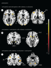
Activity during tickling and tickling in the presence of voluntary vocalization. (a) Conjunction analysis of tickling and tickling in the presence of voluntary vocalizations. Activation of the anterior (AI), posterior (PI) and limen (LI) parts of the insular cortex, of the primary sensorimotor cortex (S1/M1), the anterior cingulate cortex (ACC), the subcallosal area (SCA), the hypothalamus (HYP), the periaqueductal grey (PAG) and the brainstem lateral tegmental field in the area of the nucleus ambiguus (AMB) is shown. In the illustrated example, the level of significance for threshold activity was set at p < 0.05 (FWE-corrected). Further information concerning activation related to the nucleus accumbens and the pontine tegmentum is listed in table 1. (b) Brain activation during tickling compared with monotonous foot contacts. Activation of the anterior (AI), posterior (PI) and limen (LI) parts of the insular cortex, of the primary sensorimotor cortex (S1/M1), the ACC, the hypothalamus (HYP) and the periaqueductal grey (PAG) is shown. In the illustrated example, the level of significance for threshold activity was set at p < 0.05 (FWE-corrected). Further information concerning activation related to the middle frontal gyrus, the nucleus accumbens, the midbrain tegmentum and the anterior and the posterior cerebellar lobe is listed in table 1. (c) Brain activation during tickling in the presence of voluntary vocalizations compared with monotonous foot contacts. Activation of the primary sensorimotor cortex (S1/M1), the limen (LI) of the insular cortex, the subcallosal area (SCA) and the brainstem lateral tegmental field in the area of the nucleus ambiguus (AMB) is shown. In the illustrated example, the level of significance for threshold activity was set at p < 0.05 (FWE-corrected).
Figure 3.
Figure 3.
Activity during anticipation and anticipation in the presence of voluntary vocalization. (a) Conjunction analysis of anticipation of tickling and tickling in the presence of voluntary vocalizations. Anterior (AI) and posterior (PI) parts of the insular cortex and superior parietal lobe (SPL) are activated. In the illustrated example, the level of significance for threshold activity was set at p < 0.05 (FWE corrected). Further information concerning activation related to the sensorimotor cortex, the premotor cortex, the middle cingulate gyrus and the visual cortex is listed in table 2. (b) Brain activation during anticipation of tickling compared to anticipation of tickling in presence of voluntary vocalizations. The corpus amygdaleum (AMY), the hypothalamus (HYP) and the nucleus accumbens (NAC) are activated. In the illustrated example, the level of significance for threshold activity was set at p < 0.05 (FWE corrected). (c) Brain activation during anticipation of tickling in the presence of voluntary vocalizations compared to anticipation of tickling. The primary motor area (M1), the auditory cortex (AUD) and the anterior lobe of the cerebellum (CER) are activated. In the illustrated example, the level of significance for threshold activity was set at p < 0.05 (FWE corrected).
Figure 4.
Figure 4.
Simplified model of the efferent pathways implicated in the control of vocalization in humans (adapted from [15]). This Figure illustrates the interaction of key components of the voluntary and the involuntary pathways of vocalization. Highlighted in the centre are the regions of the brainstem that harbour the premotor interneurons and the motor neurons that control the laryngeal effectors (nucleus ambiguus) and respiratory laryngeal coordination (nucleus retroambiguus). The periaqueductal grey (PAG) is crucial for the involuntary control of vocal expression via its connections with the nucleus ambiguus and nucleus retroambiguus. Activity in the PAG is driven by that in regions associated with the limbic system, including the hypothalamus, the anterior insula and the anterior cingulate cortex (ACC). The latter is also a target of efferences of the (pre-)supplementary cortex, which is considered to bridge the voluntary and the involuntary control of vocalization [22]. The laryngeal motor cortex exerts voluntary vocal control, either directly, via connections with laryngeal effectors in the nucleus ambiguous, or indirectly, via the lateral tegmental field. Brain regions coloured in yellow represent, or bear reference to the emotional motor pathway; those in green denote the voluntary motor pathway; and those in lime green depict the effectors for vocal output.

References

    1. Sauter DA, Eisner F, Ekman P, Scott SK. 2010. Cross-cultural recognition of basic emotions through nonverbal emotional vocalizations. Proc. Natl Acad. Sci. USA 107, 2408-2412. (10.1073/pnas.0908239106) - DOI - PMC - PubMed
    1. Scheiner E, Hammerschmidt K, Jurgens U, Zwirner P. 2004. The influence of hearing impairment on preverbal emotional vocalizations of infants. Folia Phoniatr. Logopaed. 56, 27-40. (10.1159/000075326) - DOI - PubMed
    1. Roccaro-Waldmeyer DM, Babalian A, Muller A, Celio MR. 2016. Reduction in 50-kHz call-numbers and suppression of tickling-associated positive affective behaviour after lesioning of the lateral hypothalamic parvafox nucleus in rats. Behav. Brain Res. 298, 167-180. (10.1016/j.bbr.2015.11.004) - DOI - PubMed
    1. Ross MD, Owren MJ, Zimmermann E. 2009. Reconstructing the evolution of laughter in great apes and humans. Curr. Biol. 19, 1106-1111. (10.1016/j.cub.2009.05.028) - DOI - PubMed
    1. Scheiner E, Hammerschmidt K, Jurgens U, Zwimer P. 2006. Vocal expression of emotions in normally hearing and hearing-impaired infants. J. Voice 20, 585-604. (10.1016/j.jvoice.2005.09.001) - DOI - PubMed

LinkOut - more resources