Review
. 2022 Sep 20;10(1):60.
doi: 10.1038/s41413-022-00226-9.

Current understanding of osteoarthritis pathogenesis and relevant new approaches

Affiliations
Review

Current understanding of osteoarthritis pathogenesis and relevant new approaches

Liping Tong et al. Bone Res. .

Abstract

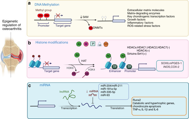
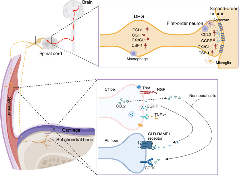
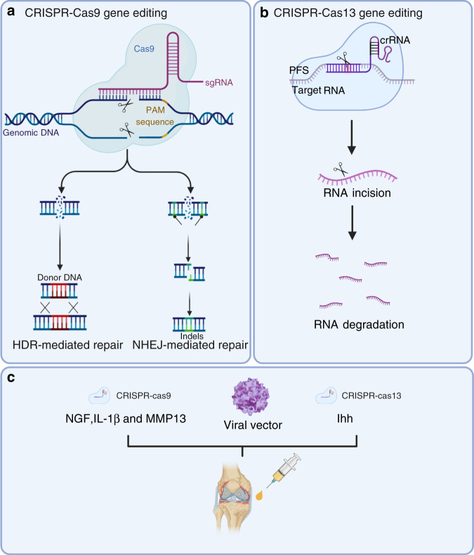
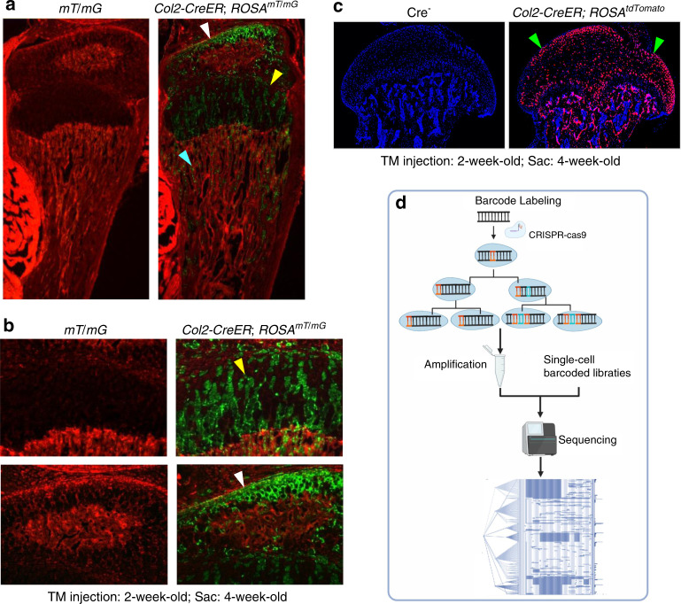
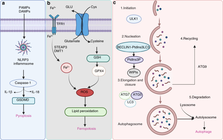
Osteoarthritis (OA) is the most common degenerative joint disease that causes painful swelling and permanent damage to the joints in the body. The molecular mechanisms of OA are currently unknown. OA is a heterogeneous disease that affects the entire joint, and multiple tissues are altered during OA development. To better understand the pathological mechanisms of OA, new approaches, methods, and techniques need to be used to understand OA pathogenesis. In this review, we first focus on the epigenetic regulation of OA, with a particular focus on DNA methylation, histone modification, and microRNA regulation, followed by a summary of several key mediators in OA-associated pain. We then introduce several innovative techniques that have been and will continue to be used in the fields of OA and OA-associated pain, such as CRISPR, scRNA sequencing, and lineage tracing. Next, we discuss the timely updates concerning cell death regulation in OA pathology, including pyroptosis, ferroptosis, and autophagy, as well as their individual roles in OA and potential molecular targets in treating OA. Finally, our review highlights new directions on the role of the synovial lymphatic system in OA. An improved understanding of OA pathogenesis will aid in the development of more specific and effective therapeutic interventions for OA.


Conflict of interest statement

The authors declare no competing interests.

Figures

Fig. 1
Fig. 1
Epigenetic regulation of osteoarthritis. Three types of epigenetic regulation of the molecular pathogenesis of OA. a DNA methylation is catalyzed by DNMTs, and abnormal changes in DNA methylation occur in the promoter regions of related genes and signaling pathways in OA chondrocytes. b Histone modifications included phosphorylation, methylation, acetylation, ubiquitination, and SUMOylation. Histone acetylation is mainly mediated by HATs, and histone deacetylation is usually catalyzed by two types of enzymes: classic histone deacetylases (HDACs) and sirtuins. Increased H3K4 methylation leads to catabolic responses mediated by iNOS and COX-2 expression in human OA chondrocytes. The methylation of H3K9 increases Sox9 and mPGES-1 expression in human OA chondrocytes. c MiR-204/miR-211, miR-181a-5p, miR-335-5p, and miR-93 inhibit Runx2, chondrocyte apoptosis, and the expression of catabolic and hypertrophic genes
Fig. 2
Fig. 2
The NGF, CGRP, CCL2/CCR2, and TNFα signaling pathways in OA-associated pain. NGF acts on its receptor TrkA and mediates pain transmission. CGRP is released from C-fiber terminals and binds with calcitonin receptor-like receptor (CLR) and receptor activity-modifying protein 1 (RAMP1) on adjacent Aδ nerve fibers. High expression of chemokines, such as CCL2 and CX3CL1, and other immune mediators, such as colony-stimulating factor 1 (CSF-1) and CGRP, was detected in the DRG and dorsal horn of the spinal cord
Fig. 3
Fig. 3
CRISPR/Cas systems have been used for gene editing and treating OA. a Cas9 generates DNA double-strand breaks that activate the DNA damage response and induce repair through various endogenous pathways: nonhomologous end joining (NHEJ) or homologous directed repair (HDR). b Cas13 cuts the target RNA via intrinsic RNase activity. c CRISPR–Cas9-mediated NGF, IL-1β, and MMP13 deletion and CRISPR–Cas13-mediated ihh knockout were used for OA treatment
Fig. 4
Fig. 4
Lineage tracing technique. ac Col2-expressing cells label a subpopulation of chondrocytes which could migrate to bone marrow underneath the growth plate and transdifferentiate into progenitor cells. a Col2-CreER mice were generated and bred with ROSAmT/mG reporter mice or ROSAtdTomato reporter mice. b Tamoxifen was intraperitoneally injected to the 2-week-old Col2-CreER; ROSAmT/mG mice or Col2-CreER; ROSAtdTomato mice for 5 consecutive days (1 mg per 10 g body weight) and mice were sacrificed at 4-week-old. c Col2-expressing cells were detected in articular chondrocytes, growth plate chondrocytes and cells located in bone marrow underneath the growth plate. d DNA barcoding technology was used for cell lineage tracing. Figure 4a, b and Fig. 4c were cited from our previous publications.
Fig. 5
Fig. 5
Pathways of pyroptosis, ferroptosis, and autophagy. a Many factors trigger assembly of the NLRP3 inflammasome, followed by activation of caspase-1 or caspase-11/4/5, which cleaves the gasdermin D (GSDMD) protein and pro-IL-1β and pro-IL-18, resulting in the release of IL-1β and IL-18. b During ferroptotic cell death, intracellular Glu is transported to the extracellular space, and extracellular Cys2 is transported into the cell, where it is then transformed into Cys for GSH synthesis. GPX4 reduces ROS accumulation. Excess iron is the basis for ferroptosis execution. Circulating iron binds with transferrin in the form of Fe3+ and then enters the cells by TFR1. Fe3+ is deoxidized to Fe2+ by the iron oxide reductase STEAP3. Ultimately, Fe2+ is released into a labile iron pool in the cytoplasm from the endosome via DMT1. c The canonical formation of autophagosomes involves the following steps: initiation, nucleation, elongation, closure, and recycling
Fig. 6
Fig. 6
Arthritogenic MMP13 is removed from osteoarthritic and inflamed joints via the synovial lymphatic system. C57BL/6J mice received sham or DMM surgery (ac), and TNF-Tg and WT control mice (df) were used. Knees and DLNs were harvested from operated mice 6 weeks post-surgery (ac) and from 6-month-old WT and TNF-Tg mice (df). a, d Frozen sections from the knees were H&E stained for light microscopy, and representative 10x images are shown (scale bar = 100 µm). F: femur, T: tibia, M: meniscus. Parallel knee sections (dotted lines indicate the joint space) and DLN sections were immunostained with green fluorescent labeled antibodies against MMP13, counterstained with DAPI, and representative dark field images obtained at 10x are shown. Note that only articular chondrocytes in PTOA knees, synovium in TNF-Tg knees, and their DLNs, have robust MMP13 immuno-reactivity. b, e VisioPharm histomorphometry was performed to quantify the percentage of MMP13+ area of the DLN immunostained sections, and data are presented for each DLN +/− SD (*P < 0.05 vs. non-arthritic control with paired t-test). c, f Mmp13 mRNA levels in knees were assessed via qPCR. The data are presented for each tissue ± SD (*P < 0.05 vs. non-arthritic control with paired t-test). The content presented in Fig. 6 was derived from a PhD dissertation and was provided by Dr. Xi Lin, with her permission, a postdoctoral fellow in University of Rochester Medical Center

References

    1. Chen D. Osteoarthritis: a complicated joint disease requiring extensive studies with multiple approaches. J. Orthop. Translat. 2022;32:130. doi: 10.1016/j.jot.2022.02.009. - DOI - PMC - PubMed
    1. Chen D, et al. Osteoarthritis: toward a comprehensive understanding of pathological mechanism. Bone Res. 2017;5:16044. doi: 10.1038/boneres.2016.44. - DOI - PMC - PubMed
    1. Li Y, Xie W, Xiao W, Dou D. Progress in osteoarthritis research by the National Natural Science Foundation of China. Bone Res. 2022;10:41. doi: 10.1038/s41413-022-00207-y. - DOI - PMC - PubMed
    1. Sun AR, et al. Cartilage tissue engineering for obesity-induced osteoarthritis: physiology, challenges, and future prospects. J. Orthop. Translat. 2021;26:3–15. doi: 10.1016/j.jot.2020.07.004. - DOI - PMC - PubMed
    1. Wu XH, Lai YM, Cao HL, Xiao GZ. Kindlin-2 preserves integrity of the articular cartilage to protect against osteoarthritis. J. Bone Min. Res. 2022;37:45–46. doi: 10.1002/jbmr.4499. - DOI - PubMed