. 2022 Sep 20;13(1):5518.
doi: 10.1038/s41467-022-33254-4.

Smart soft contact lenses for continuous 24-hour monitoring of intraocular pressure in glaucoma care

Affiliations

Smart soft contact lenses for continuous 24-hour monitoring of intraocular pressure in glaucoma care

Jinyuan Zhang et al. Nat Commun. .

Abstract

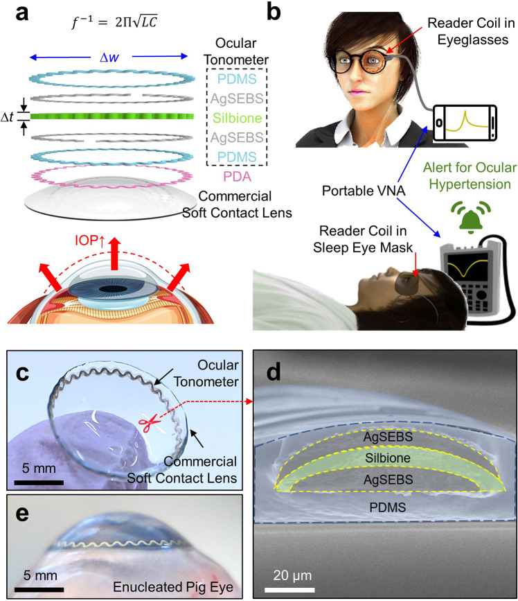
Continuous monitoring of intraocular pressure, particularly during sleep, remains a grand challenge in glaucoma care. Here we introduce a class of smart soft contact lenses, enabling the continuous 24-hour monitoring of intraocular pressure, even during sleep. Uniquely, the smart soft contact lenses are built upon various commercial brands of soft contact lenses without altering their intrinsic properties such as lens power, biocompatibility, softness, transparency, wettability, oxygen transmissibility, and overnight wearability. We show that the smart soft contact lenses can seamlessly fit across different corneal curvatures and thicknesses in human eyes and therefore accurately measure absolute intraocular pressure under ambulatory conditions. We perform a comprehensive set of in vivo evaluations in rabbit, dog, and human eyes from normal to hypertension to confirm the superior measurement accuracy, within-subject repeatability, and user comfort of the smart soft contact lenses beyond current wearable ocular tonometers. We envision that the smart soft contact lenses will be effective in glaucoma care.


Conflict of interest statement

The authors declare no competing interests.

Figures

Fig. 1
Fig. 1. Schematics and optical images of the SSCL.
a Layered schematic view of the SSCL. b Data acquisition scheme for the monitoring of daytime (top panel) and nighttime (bottom panel) IOP. c Photograph of the SSCL. d Cross-sectional SEM image of the SSCL. e Photograph of the SSCL in an enucleated pig eye.
Fig. 2
Fig. 2. Benchtop evaluations.
a Stress–strain curve for the SSCL (red line), the bare soft contact lens without the ocular tonometer (blue line), and the ocular tonometer without the soft contact lens (green line). b Resistance response (ΔR/R0) of the SSCL following 10,000 cycles of stretching at 25% (blue line) and 50% (red line). c Capacitance response (ΔC/C0) of the SSCL with respect to applied pressure by employing three different dielectric interlayers made of Silbione (red line), Ecoflex (blue line), and PDMS (green line) in the ocular tonometer. d Normalized baseline resonant frequency of the SSCL following 1000 cycles of flipping, rubbing, folding, and stretching. e Five cycles of disinfecting (red line) and cleaning (blue line); f 5 cycles of hydrating (red line) and dehydrating (blue line); g 5 cycles of heating and cooling; and h soaking in a saline solution for 30 days. i Adhesive strength of the PDA layer after multiple cycles of handling, disinfecting, dehydrating, and heating. The error bars represent standard deviations with n = 5 for each group.
Fig. 3
Fig. 3. Ex vivo evaluations in enucleated pig eyes.
a Schematic view of the ex vivo measurement setup. b Photograph of the SSCL in an enucleated pig eye during measurement. c Reflection spectra (S11) of the SSCL in response to ascending and descending IOP of the pig eye. d Resonant frequency of the SSCL in the response to the IOP of enucleated pig eyes (n = 3). e Baseline resonant frequency of the SSCL at various angles with respect to the reader coil (n = 5). f Baseline resonant frequency of the SSCL at various distances with respect to the reader coil (n = 5).
Fig. 4
Fig. 4. Cell viability and in vivo evaluations in rabbit eyes.
a Cell viability assay of HCEpiCs seeded on the SSCL without (blue bars) and with (green bars) the presence of the PDA adhesive as compared to the bare soft contact lens (red bars) (n = 4). b Quantified accumulation of proteins on the bare soft contact lens (red bar) and the SSCL (blue bar) before (left column, p = 0.02194 before disinfecting the bare soft contact lens vs. the ocular tonometer, n = 5; p = 0.00115 before vs. after disinfecting the bare soft contact lens, n = 5) and after (right column, p = 0.00979 before vs. after disinfecting the ocular tonometer, n = 5) disinfection. Significance was set at ***p < 0.01, and **p < 0.1. c Representative surface fluorescence image of the bare soft contact lens (top row) and the SSCL (bottom row) before (left column) and after (right column) disinfection. d Photographs of the rabbit eye wearing the SSCL (left panel) and the bare soft contact lens (right panel). e Representative AS-OCT image of the rabbit eye after 1 h of wearing the SSCL. f Representative photomicrographs of the rabbit eye with H&E staining displaying epithelial erosion and mild mixed inflammation with edema at the limbus after 24-h wear of the SSCL (left panel) and the bare soft contact lens (right panel). g Representative photomicrographs of the rabbit eye with H&E staining displaying no abnormality at the limbus after 2-week wear of the SSCL (left panel) as compared to those of the naked eye (right panel).
Fig. 5
Fig. 5. In vivo sensing performance in dog eyes.
a Photographs of a dog wearing the SSCL and a dog goggle (V2 Goggle; Rex Specs, Inc.) embedded with the reader coil. b Resonant frequency of the SSCL with respect to the IOP values of the dog (n = 3). c 24-h IOP rhythm of the dog obtained from the SSCL (blue line) and the iCare Tonovet Plus (red line) (n = 3).
Fig. 6
Fig. 6. In-clinic examinations in human eyes.
a Photograph (left panel) and slit lamp biomicroscopic image (right panel) of a human eye wearing the SSCL. b Representative AS-OCT image of the eye after 1 h of wearing the SSCL. c Representative visual field of the eye wearing the SSCL (top panel) and the naked eye (bottom panel). d Representative slit lamp fluorescent images of the eye after 6 h of wearing the SSCL (top panel) and the naked eye (bottom panel). e Average user comfort rating for the SSCL (red line), the bare soft contact lens (blue line), and the Triggerfish lens (green line) from three participants.
Fig. 7
Fig. 7. Ambulatory IOP monitoring in human eyes.
a Photograph of a participant wearing the SSCL in a sitting posture with the reader coil embedded within a typical eyeglass frame and b a sleep eye mask. c Reflection spectra (S11) of the SSCL in response to various body postures. d Resonant frequency of the SSCL in each body posture for three participants with different corneal curvatures and thicknesses (n = 3). e Calibrated absolute IOP data of a participant using the SSCL (red line), the iCare Home (green line), and the Triggerfish lens (blue line). f Time-varying change in the ambulatory IOP of a participant obtained using the SSCL (red line), the GAT (blue line), and the iCare Home (green line) for 6 h (n = 3).

References

    1. Weinreb RN, Aung T, Medeiros FA. The pathophysiology and treatment of glaucoma: a review. JAMA. 2014;311:1901–1911. doi: 10.1001/jama.2014.3192. - DOI - PMC - PubMed
    1. Beykin G, Norcia AM, Srinivasan VJ, Dubra A, Goldberg JL. Discovery and clinical translation of novel glaucoma biomarkers. Prog. Retin. Eye Res. 2021;80:100875. doi: 10.1016/j.preteyeres.2020.100875. - DOI - PMC - PubMed
    1. Garway-Heath DF, et al. Latanoprost for open-angle glaucoma: a randomised, multicentre, placebo-controlled trial. Lancet. 2015;385:1295–1304. doi: 10.1016/S0140-6736(14)62111-5. - DOI - PubMed
    1. Li G, et al. In vivo measurement of trabecular meshwork stiffness in a corticosteroid-induced ocular hypertensive mouse model. Proc. Natl Acad. Sci. USA. 2019;116:1714–1722. doi: 10.1073/pnas.1814889116. - DOI - PMC - PubMed
    1. Gordon MO, Kass MA. What we have learned from the ocular hypertension treatment study? Am. J. Ophthalmol. 2018;189:xxiv–xxvii. doi: 10.1016/j.ajo.2018.02.016. - DOI - PMC - PubMed

Publication types