. 2023 Apr;28(4):1813-1826.
doi: 10.1038/s41380-022-01769-3. Epub 2022 Sep 21.

Mitochondrial DNA variation in Alzheimer's disease reveals a unique microprotein called SHMOOSE

Affiliations

Mitochondrial DNA variation in Alzheimer's disease reveals a unique microprotein called SHMOOSE

Brendan Miller et al. Mol Psychiatry. 2023 Apr.

Erratum in

  • Correction: Mitochondrial DNA variation in Alzheimer's disease reveals a unique microprotein called SHMOOSE.
    Miller B, Kim SJ, Mehta HH, Cao K, Kumagai H, Thumaty N, Leelaprachakul N, Braniff RG, Jiao H, Vaughan J, Diedrich J, Saghatelian A, Arpawong TE, Crimmins EM, Ertekin-Taner N, Tubi MA, Hare ET, Braskie MN, Décarie-Spain L, Kanoski SE, Grodstein F, Bennett DA, Zhao L, Toga AW, Wan J, Yen K, Cohen P; Alzheimer’s Disease Neuroimaging Initiative. Miller B, et al. Mol Psychiatry. 2023 Apr;28(4):1827. doi: 10.1038/s41380-023-01956-w. Mol Psychiatry. 2023. PMID: 36658336 No abstract available.

Abstract

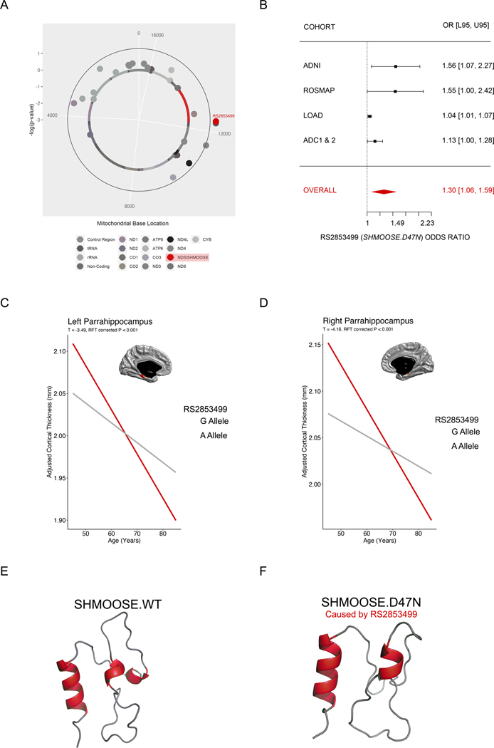
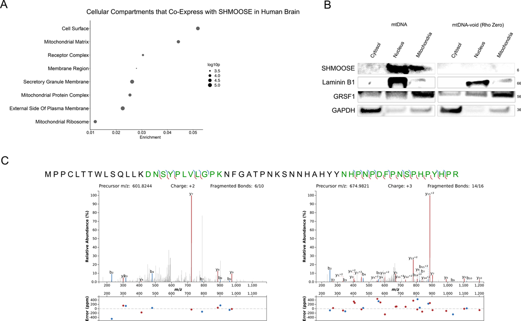
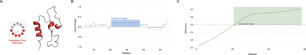
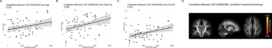
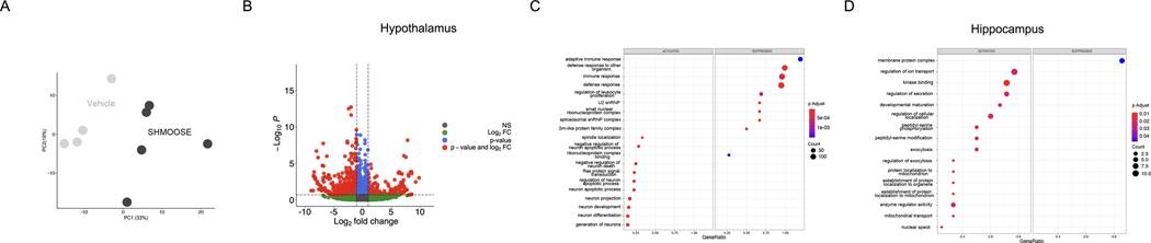
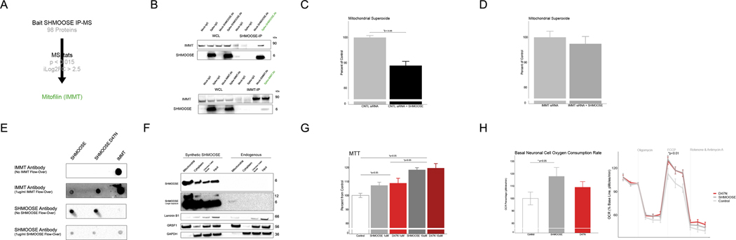
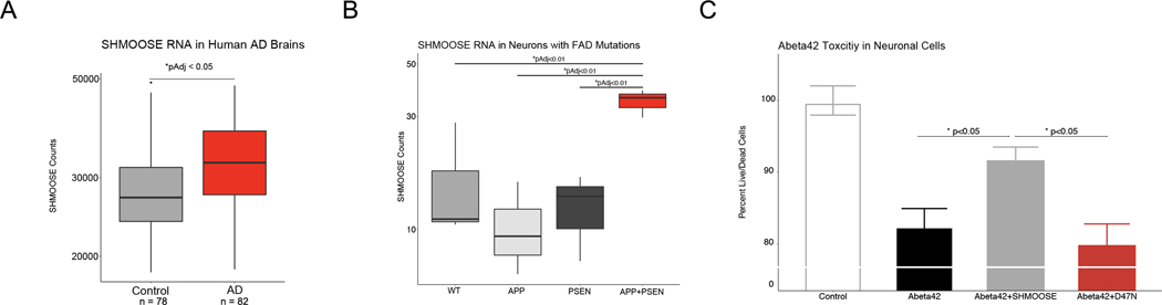
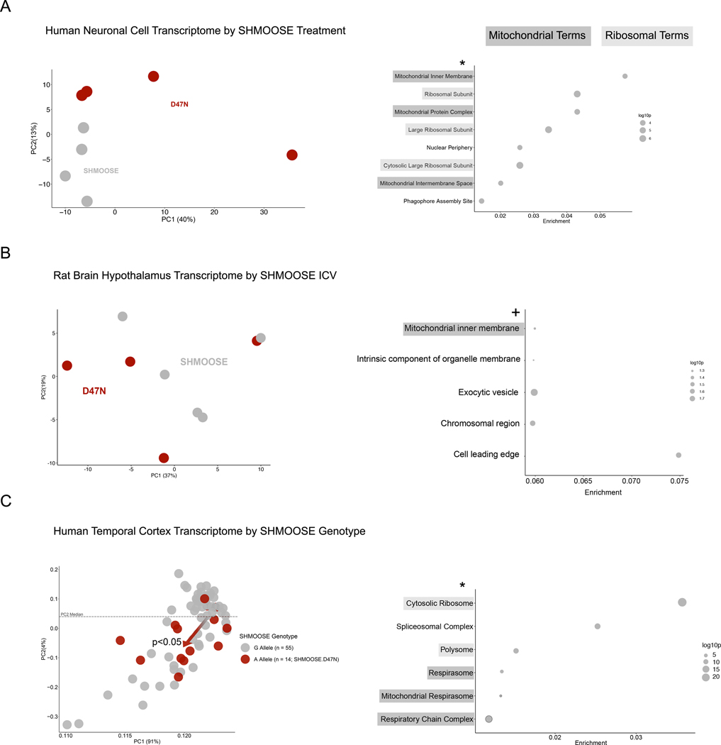
Mitochondrial DNA variants have previously associated with disease, but the underlying mechanisms have been largely elusive. Here, we report that mitochondrial SNP rs2853499 associated with Alzheimer's disease (AD), neuroimaging, and transcriptomics. We mapped rs2853499 to a novel mitochondrial small open reading frame called SHMOOSE with microprotein encoding potential. Indeed, we detected two unique SHMOOSE-derived peptide fragments in mitochondria by using mass spectrometry-the first unique mass spectrometry-based detection of a mitochondrial-encoded microprotein to date. Furthermore, cerebrospinal fluid (CSF) SHMOOSE levels in humans correlated with age, CSF tau, and brain white matter volume. We followed up on these genetic and biochemical findings by carrying out a series of functional experiments. SHMOOSE acted on the brain following intracerebroventricular administration, differentiated mitochondrial gene expression in multiple models, localized to mitochondria, bound the inner mitochondrial membrane protein mitofilin, and boosted mitochondrial oxygen consumption. Altogether, SHMOOSE has vast implications for the fields of neurobiology, Alzheimer's disease, and microproteins.


Conflict of interest statement

Competing Interests

Intellectual property related to SHMOOSE has been filed by the University of Southern California.

Figures

Fig. 1.
Fig. 1.. Mitochondrial rs2853499 predicts AD risk and parahippocampus thickness.
A The GWAS Manhattan plot-equivalent of a MiWAS, called a Solar Plot. SNPs extending beyond the outer blue are statistically significant by a permutation empirical p value of 0.05. The most significant mtSNPs were rs2853498 and rs2853499 – both are haplogroup U determining, with the latter causing the missense change to SHMOOSE. B Meta-analysis forest plot of rs2853499 in ADNI, ROSMAP, LOAD, and ADC/1/2. Bars represent 95% confidence intervals. C Neuroimaging-based PheWAS in UK Biobank that illustrates the significant effects of SHMOOSE.D47N and age on the left parahippocampus. D Neuroimaging-based PheWAS in UK Biobank that illustrates the significant effects of SHMOOSE.D47N and age on the right parahippocampus E RosettaTTFold prediction of SHMOOSE. F RosettaTTFold prediction of SHMOOSE.D47N, caused by rs2853499.
Fig. 2.
Fig. 2.. Endogenous SHMOOSE is detected in mitochondria.
A GO cellular terms enriched by genes that co-express with SHMOOSE in the human temporal cortex (n = 69). B Western blot detection of ~6kDa SHMOOSE in cells containing mtDNA versus cells not containing mtDNA (i.e., rho zero cells). Laminin B1 is a nuclear marker; GRSF1 isoform is a mitochondrial marker; GAPDH is a cytosolic marker. C Unique mass spectrometry SHMOOSE-derived fragments detected in mitochondria fractions.
Fig. 3.
Fig. 3.. Unique features of the SHMOOSE amino acid sequence.
A 3–11 amphipathic prediction output from HeliQuest that illustrates a potential hydrophobic face (red) derived from the RosettaFold structure model. B Positively charged domain of SHMOOSE that is highlighted in blue. C Disordered protein region that is highlighted in green as predicted by IUPred3.
Fig. 4.
Fig. 4.. SHMOOSE levels correlate with AD-related biomarkers and brain white matter.
A Human CSF SHMOOSE levels (pg/ml) correlation with age. Regression model includes biological sex as a covariate; p value < 0.001. B SHMOOSE correlation with CSF total tau (pg/ml). Regression model includes biological sex and age as covariates; p value < 0.05. C SHMOOSE correlation with CSF phosphorylated tau at residue 181 (p tau 181; pg/ml). Regression model includes biological sex and age as covariates; p value < 0.05. D Higher CSF SHMOOSE was significantly associated with lower DTI FA in the body of the corpus callosum and bilateral superior corona radiata in 72 non-demented older adults. Regression model included age, reported sex, and clinical dementia rating score. Colored voxels indicate FSLThreshold-Free Cluster Enhancement-derived p values < 0.05 after correction for voxelwise multiple comparisons. Presented in radiological orientation (L=R).
Fig. 5.
Fig. 5.. Direct actions of SHMOOSE on brain gene expression.
A. Principal component analysis for vehicle and SHMOOSE on hypothalamic gene expression following ICV administration. B Corresponding volcano plot that is color coded by significance (blue or red) and log fold change significance (red). C GSEA significant terms that were enriched by SHMOOSE-induced gene expression in the hypothalamus. D GSEA significant terms that were enriched by SHMOOSE-induced gene expression in the hippocampus.
Fig. 6.
Fig. 6.. SHMOOSE interacts with mitofilin and modifies mitochondrial biology.
A Schematic proteomics analysis of SHMOOSE-spiked neural cell lysates that were immunoprecipitated using a custom SHMOOSE polyclonal antibody. B Reciprocal western blot validation of SHMOOSE/mitofilin interaction from SHMOOSE-treated cells. C SHMOOSE lowered mitochondrial superoxide levels, D yet SHMOOSE had no effect when mitofilin was knocked down with siRNA. Y axis is normalized to control baseline values. E SHMOOSE-treated differentiated SH-SY5Y cells (1 uM) localized to mitochondria after 15 minutes. Top portion of the blot represents a 5-second exposure. Second portion of the blot represents a 30-second exposure. SHMOOSE dimerized around 12 kDa in the SHMOOSE-treated conditions. Laminin, GRSF1, and GAPDH represent nuclear, mitochondrial, and cytosolic fractions, respectively. F Reciprocal dot blot illustrating recombinant mitofilin and SHMOOSE and SHMOOSE.D47N interact. G 1uM of both SHMOOSE (gray) and SHMOOSE.D47N (red) both increased metabolic activity (MTT) at 1uM and 10uM. Significant bars represent differences among dosage groups. H Normalized to the baseline third measurement, SHMOOSE increased oxygen consumption rate. SHMOOSE and SHMOOSE.D47N increased mitochondrial spare capacity. Significance defined as p value < 0.05 for independent t tests.
Fig. 7.
Fig. 7.. SHMOOSE expression in human Alzheimer’s disease brains and activity in neuronal models of amyloid beta toxicity.
A SHMOOSE RNA expression in the temporal cortex of AD cases (red). Significance represented as pAdj < 0.05 following negative binomial regression on all normalized mitochondrial gene counts. B SHMOOSE expression in neurons derived from iPSCs with FAD APP mutation (light grey), FAD PSEN mutation (dark grey), and FAD APP plus PSEN mutations (red). SHMOOSE expression was highest in the latter cell type. Significance defined as pAdj < 0.05 following negative binomial regression on all normalized mitochondrial gene counts. C SHMOOSE-treated neural cells protects against amyloid beta 42-induced toxicity (grey). Significance defined as p value < 0.05 for independent t test.
Fig. 8.
Fig. 8.. Bulk gene expression differences between SHMOOSE and mutant D47N indicate altered function
Dark grey highlighting represents mitochondrial terms and light grey highlighting represents ribosomal terms. A Principal component analysis (PCA) of the in vitro gene expression signature for 10 uM SHMOOSE or SHMOOSE.D47N-treated neural cells after 24 hours. To the right of the PCA figure are GO cellular compartment terms enriched by SHMOOSE.D47N-treated neural cells, and the * indicates statistical significance following FDR. B PCA of the rat ICV gene expression signature for SHMOOSE or SHMOOSE.D47N after 24 hours. To the right of the PCA figure are GO cellular compartments that were nominally significant, and the + indicates nominal significance prior to FDR. C PCA for 14 SHMOOSE.D47N carriers (red) or 55 SHMOOSE reference allele carriers (grey). Dashed line represents the median value of PC2. Of the 14 SHMOOSE.D47N carriers, 11 fall below the median PC2 value (p value < 0.05; generalized linear model), which is represented by the red arrow at the center of the reference cluster. To the right of the PCA figure are GO cellular compartment terms significantly enriched by SHMOOSE.D47N carriers, and the * indicates statistical significance following FDR.

References

    1. Saghatelian A, Couso JP. Discovery and characterization of smORF-encoded bioactive polypeptides. Nat Chem Biol 2015; 11(12): 909–916. - PMC - PubMed
    1. Martinez TF, Chu Q, Donaldson C, Tan D, Shokhirev MN, Saghatelian A. Accurate annotation of human protein-coding small open reading frames. Nat Chem Biol 2020; 16(4): 458–468. - PMC - PubMed
    1. Mudge JM, Ruiz-Orera J, Prensner JR, Brunet MA, Calvet F, Jungreis I et al. Standardized annotation of translated open reading frames. Nat Biotechnol 2022; 40(7): 994–999. - PMC - PubMed
    1. Miller B, Kim SJ, Kumagai H, Mehta HH, Xiang W, Liu J et al. Peptides derived from small mitochondrial open reading frames: Genomic, biological, and therapeutic implications. Exp Cell Res 2020: 112056. - PMC - PubMed
    1. Kim SJ, Guerrero N, Wassef G, Xiao J, Mehta HH, Cohen P et al. The mitochondrial-derived peptide humanin activates the ERK1/2, AKT, and STAT3 signaling pathways and has age-dependent signaling differences in the hippocampus. Oncotarget 2016; 7(30): 46899–46912. - PMC - PubMed

Publication types