. 2022 Nov;19(11):1251-1262.
doi: 10.1038/s41423-022-00919-5. Epub 2022 Sep 20.

Macrophage-specific inhibition of the histone demethylase JMJD3 decreases STING and pathologic inflammation in diabetic wound repair

Affiliations

Macrophage-specific inhibition of the histone demethylase JMJD3 decreases STING and pathologic inflammation in diabetic wound repair

Christopher O Audu et al. Cell Mol Immunol. 2022 Nov.

Abstract

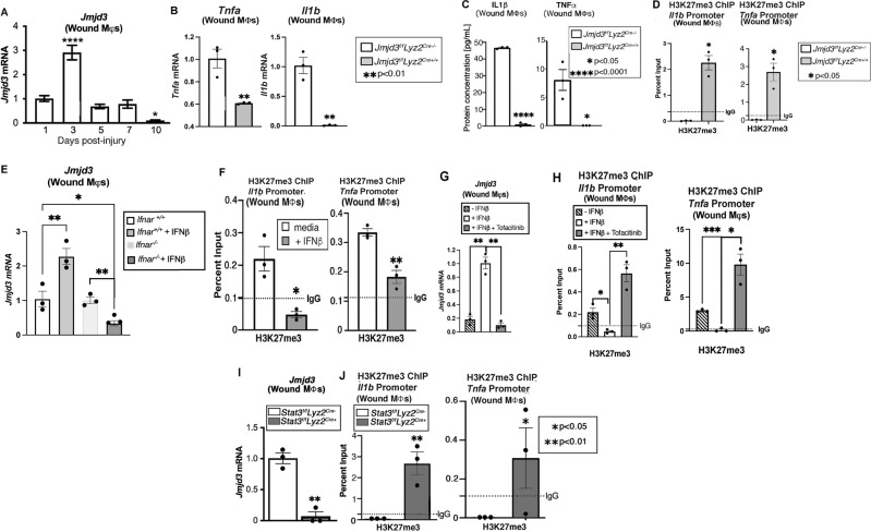
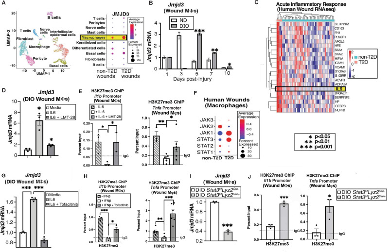
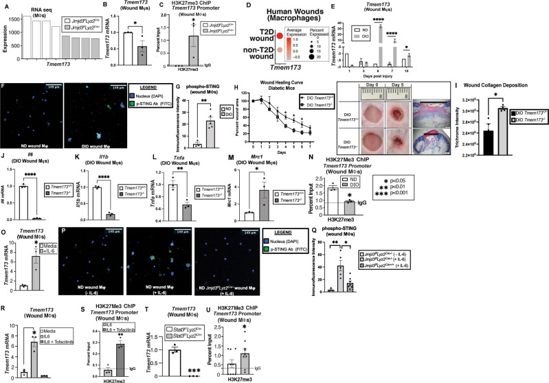
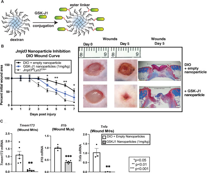
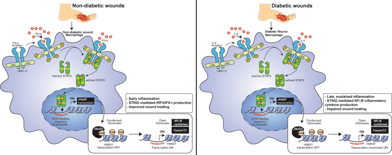
Macrophage plasticity is critical for normal tissue repair following injury. In pathologic states such as diabetes, macrophage plasticity is impaired, and macrophages remain in a persistent proinflammatory state; however, the reasons for this are unknown. Here, using single-cell RNA sequencing of human diabetic wounds, we identified increased JMJD3 in diabetic wound macrophages, resulting in increased inflammatory gene expression. Mechanistically, we report that in wound healing, JMJD3 directs early macrophage-mediated inflammation via JAK1,3/STAT3 signaling. However, in the diabetic state, we found that IL-6, a cytokine increased in diabetic wound tissue at later time points post-injury, regulates JMJD3 expression in diabetic wound macrophages via the JAK1,3/STAT3 pathway and that this late increase in JMJD3 induces NFκB-mediated inflammatory gene transcription in wound macrophages via an H3K27me3 mechanism. Interestingly, RNA sequencing of wound macrophages isolated from mice with JMJD3-deficient myeloid cells (Jmjd3f/fLyz2Cre+) identified that the STING gene (Tmem173) is regulated by JMJD3 in wound macrophages. STING limits inflammatory cytokine production by wound macrophages during healing. However, in diabetic mice, its role changes to limit wound repair and enhance inflammation. This finding is important since STING is associated with chronic inflammation, and we found STING to be elevated in human and murine diabetic wound macrophages at late time points. Finally, we demonstrate that macrophage-specific, nanoparticle inhibition of JMJD3 in diabetic wounds significantly improves diabetic wound repair by decreasing inflammatory cytokines and STING. Taken together, this work highlights the central role of JMJD3 in tissue repair and identifies cell-specific targeting as a viable therapeutic strategy for nonhealing diabetic wounds.

Keywords: JMJD3; STING; diabetes; epigenetics; wound healing.


Conflict of interest statement

The authors declare no competing interests.

Figures

Fig. 1
Fig. 1
Jmjd3 directs early inflammation in nondiabetic wound Mφs via the JAK1/3/STAT3 pathway. A Jmjd3 expression from wound Mφs (CD3-/CD19-/NK1.1-/Ly6G-/CD11b+) harvested on Days 1–10 after wounding (6 mm punch biopsy; N = 4/group, repeated two times). B Tnfa and Il1b expression measured in wound Mφs isolated on Day 3 post-injury from Jmjd3f/fLyz2Cre+ mice and littermate controls (N = 5/group, pooled and repeated in triplicate). C Protein levels of TNF-α and IL-1β analyzed by ELISAs from Jmjd3f/fLyz2Cre+ wound Mφs and controls isolated on Day 3 post-injury (N = 5/group, pooled and repeated in triplicate). D Wound Mφs from Jmjd3f/fLyz2Cre+ mice and littermate controls were isolated on Day 3, and chromatin immunoprecipitation (ChIP) analysis for H3K7me3 on the Tnfa and Il1b promoters was performed compared to the IgG control (dotted line). (N = 4/group, pooled and repeated in triplicate). E Wound Mφs were isolated on Day 3 post-injury from Ifnar−/− mice and controls (Ifnar+/+) and stimulated ex vivo with IFN-β (100 U; 8.5 ng/mL) for 6 h, and Jmjd3 expression was analyzed by RT‒PCR (N = 3–5/group, pooled, repeated in triplicate). F ChIP analysis of H3K27me3 at the Il1b and Tnfa promoters from Day 3 isolated wound Mφs following ex vivo IFN-β stimulation (100 U; 8.5 ng/mL) for 6 h. G Jmjd3 expression in wound Mφs following ex vivo IFN-β stimulation (100 U; 8.5 ng/mL) with and without JAK1,3 inhibition with tofacitinib (100 μM; 6 hr incubation, N = 5/group, pooled, repeated in triplicate). H ChIP analysis for H3K27me3 at the Il1b and Tnfa promoters in wound Mφs following ex vivo IFN-β stimulation (100 U; 8.5 ng/mL) with and without JAK1,3 inhibition with tofacitinib (100 μM; 6 hr incubation, N = 5/group, pooled, repeated in triplicate). I Jmjd3 expression in Stat3f/fLyz2Cre+ mice and littermate control wound Mφs following ex vivo IFN-β stimulation (100 U; 8.5 ng/mL, N = 5/group, repeated in triplicate). H ChIP analysis of H3K27me3 at the Il1b and Tnfa promoters in Stat3f/fLyz2Cre+ wound Mφs following ex vivo IFN-β stimulation (100 U; 8.5 ng/mL) for 6 h (N = 5/group, pooled, repeated in triplicate). *p < 0.05, **p < 0.01, ***p < 0.001, ****p < 0.0001. Data are presented as the mean ± SEM. Data were first analyzed for a normal distribution, and if the data passed the normality test, a two-tailed Student’s t test was used
Fig. 2
Fig. 2
Jmjd3 increases late in diabetic wound Mφs. A Cluster analysis UMAP of single-cell RNA sequencing from human T2D and non-T2D wounds showed 10 unique cell clusters (representative). Dot plot demonstrating JMJD3 expression within the Mφ population in human T2D and non-T2D wound samples (N = 42). The dot size corresponds to the proportion of cells within the group expressing each transcript, while the dot color corresponds to the expression level. B DIO and ND wound Mφs (CD3-/CD19-/NK1.1-/Ly6G-/CD11b+) were harvested on Days 1–10 and analyzed for Jmjd3 expression by RT‒PCR (N = 4/group, repeated once). C Human bulk RNA sequencing heatmap reflecting the expression profiles for selective genes (rows) across different samples (columns; stratified by T2D status) from acute inflammatory response Gene Ontology pathway analysis with upregulation of IL-6 in T2D wounds compared to control wounds (N = 42). D DIO wound Mφs were isolated on Day 5, treated ex vivo for 6 h with recombinant IL-6 (rIL-6; 20 nM) with and without IL-6 receptor inhibition (LMT-28; 200 nM) and analyzed for Jmjd3 expression (N = 3–5/group, pooled, repeated in triplicate). E ChIP analysis for H3K27me3 on the Tnfa and Il1b promoters from diabetic wound Mφs with and without IL-6 receptor inhibition (LMT-28; 200 nM; N = 3–5/group, pooled and repeated in triplicate). F Human single-cell RNA sequencing dot plot demonstrating Jak/Stat gene expression within the Mφ population in human T2D and non-T2D wound samples. (Cluster analysis UMAP shown above in (A)). The dot size corresponds to the proportion of cells within the group expressing each transcript, while the dot color corresponds to the expression level. G Jmjd3 expression following rIL-6 stimulation (20 nM) and Jak1/3 inhibition (tofacitinib; 100 μM) in diabetic wound Mφs isolated from Day 5 wounds and treated ex vivo for 4 h. (N = 5/group, pooled and repeated in triplicate). H ChIP analysis for H3K27me3 on the Tnfa and Il1b promoters from diabetic wound Mφs following ex vivo rIL-6 stimulation (20 nM) with and without JAK1/3 inhibition (tofacitinib; 100 μM) for 4 h (N = 5/group, pooled, repeated in triplicate). I Jmjd3 expression in DIO Stat3f/fLyz2Cre+ and DIO littermate control wound Mφs following ex vivo rIL-6 stimulation (20 nM) for 6 h. (N = 3/group, pooled, repeated in triplicate). J ChIP analysis for H3K27me3 on Tnfa and Il1b promoters from DIO Stat3f/fLyz2Cre+ and DIO littermate control wound Mφs following rIL-6 stimulation (20 nM) for 6 h (N = 4/group, pooled, repeated in triplicate). Data are presented as the mean ± SEM. All data are representative of 2–4 independent experiments
Fig. 3
Fig. 3
STING-mediated inflammation is regulated by JMDJ3 in wound Mφs. A Bulk RNA sequencing analysis of wound Mφs (CD3-/CD19-/NK1.1-/Ly6G-/CD11b+) isolated on Day 5 from Jmjd3f/fLyz2Cre+ mice and littermate controls with Tmem173 gene expression (N = 4/group). B, C Tmem173 expression and ChIP analysis of H3K27me3 at the Tmem173 promoter in Jmjd3f/fLyz2Cre+ and littermate control wound Mφs (N = 4/group, pooled, repeated in triplicate). D Human single-cell RNA sequencing dot plot demonstrating Tmem173 gene expression within the Mφ population in human T2D and non-T2D wound samples. Cluster analysis UMAP shown above in (2A). The dot size corresponds to the proportion of cells within the group expressing each transcript, while the dot color corresponds to the expression level. E Tmem173 gene expression from murine diabetic wound Mφs harvested on Days 1–10 compared with that of the nondiabetic controls (N = 4/group, repeated once). F Immunofluorescence against phospho-STING antibody (FITC) in ND and DIO wound Mφs. G Quantification of the immunofluorescence intensity of phospho-STING (N = 3–5 cells/group). H Wound healing curve for DIO STING knockout (DIO Tmem173−/−) mice and littermate controls (DIO Tmem173+/+), with representative healing images on Days 0 and 5 post-injury (6 mm punch wounds; 3–4 mice/group; repeated once). Wounds were harvested on Day 5, paraffin embedded, and stained with Masson’s trichrome stain (N = 3 mice/group). Representative images are shown at ×2 magnification. The black horizontal bar above the wound represents the entire wound distance, the epithelial tongues are denoted by arrowheads, and the asterisk (*) denotes wound debris. The scale bar represents 500 μm. I Collagen quantification of trichrome staining in DIO Tmem173+/+ and DIO Tmem173−/− mice (N = 4 wounds/strain; repeated once). J Il6 mRNA expression in Day 5 wound Mφs from DIO Tmem173+/+ and DIO Tmem173-/- mice. (N = 4 mice/group, pooled, repeated in triplicate). K Il1b mRNA expression in Day 5 wound Mφs from DIO Tmem173+/+ and DIO Tmem173−/− mice. (N = 4 mice/group, pooled, repeated in triplicate). L Tnfa mRNA expression in Day 5 wound Mφs from DIO STING+/+ and DIO STING−/− mice. (N = 4 mice/group, pooled, repeated in triplicate). M Mrc1 mRNA expression in Day 5 wound Mφs from DIO Tmem173+/+ and DIO Tmem173−/− mice. (N = 4 mice/group, pooled, repeated in triplicate). N ChIP analysis of H3K27me3 at the Tmem173 gene promoter in ND and DIO wound Mφs (N = 4/group, pooled, repeated in triplicate). O Tmem173 expression in wound Mφs following rIL-6 stimulation (20 nM) for 4 h (N = 5/group, pooled, repeated in triplicate). P Immunofluorescence against phospho-STING antibody (FITC) in ND wound Mφs ± rIL-6 stimulation (20 nM; 1 hr) and in Jmjd3f/fLyz2Cre+ wound Mφs treated with rIL-6 (20 nM; 1 h). Q Quantification of the immunofluorescence intensity of phospho-STING (FITC) by ImageJ (NIH) (N = 3–5 cells/group). R Tmem173 expression in wound Mφs following rIL-6 stimulation (20 nM) for 6 h with and without JAK1/3 inhibition (tofacitinib; 100 μM; N = 3/group, pooled, repeated in triplicate). S ChIP analysis of H3K27me3 on the Tmem173 promoter in wound Mφs following rIL-6 stimulation (20 nM) for 4 h with and without Jak1/3 inhibition (tofacitinib; 100 μM, N = 4/group, pooled, repeated in triplicate). T Tmem173 expression from Stat3f/fLyz2Cre+ and littermate control wound Mφ following rIL-6 stimulation (20 nM) for 4 h (N = 3/group, pooled, repeated in triplicate). U ChIP analysis of H3K27me3 on the Tmem173 gene promoter from Stat3f/fLyz2Cre+ and littermate control wound Mφs following rIL-6 stimulation (20 nM) for 4 h (N = 6/group, pooled, repeated in triplicate). *p < 0.05, **p < 0.01, ***p < 0.001. Data are presented as the mean ± SEM
Fig. 4
Fig. 4
Mφ-specific nanotherapy against JMJD3 improves diabetic tissue repair. A Schematic of Mφ specific, GSK-J1-laden, dextran core nanoparticles. B Wound healing curve for DIO mice wounded with a 6 mm punch biopsy, and wounds were injected daily starting on Day 1 post-injury with nanoparticles containing either a selective JMJD3 inhibitor (GSK-J1; 1 mg/kg) or dextrose control. DIO mice deficient in myeloid JMJD3 production (Jmjd3f/fLyz2Cre+) were included as a control. Wound area was measured daily with ImageJ software throughout the wound healing course (N = 5/group, repeated twice). Representative wound images at ×2 magnification on Day 0 and Day 5 are shown. Wounds were harvested on Day 5, paraffin embedded, and stained with Masson’s trichrome stain (N = 5–6 mice/group). The black bar above the wound represents the entire wound distance, the arrowheads represent the epithelial tongue edges, and the asterisk (*) represents wound debris. The scale bar represents 200 μm. C Tmem173, Il1b and Tnfa expression in DIO wound Mφs (CD3-/CD19-/NK1.1-/Ly6G-/CD11b+) harvested on Day 5 from wounds treated with and without GSK-J1 nanoparticles (N = 3–4/group, pooled, repeated in triplicate). *p < 0.05, **p < 0.01. Data are presented as the mean ± SEM. Data were first analyzed for normal distribution, and if data passed the normality test, a two-tailed Student’s t test was used
Fig. 5
Fig. 5
Schematic of the mechanism of JMJD3 activity in wound Mφs. (Left) Nondiabetic wound Mφs are stimulated by IFN-β, which acts through the JAK/STAT3 pathway to stimulate the early transcription of JMJD3 and facilitate early inflammation. This cascade primes STING toward a TBK1/IRF3/IFN-I pathway and ultimately leads to wound repair. (Right) In diabetic wounds, Mφs are stimulated by IL-6 through a JAK/STAT3 pathway to stimulate the late transcription of JMJD3, leading to late, sustained inflammation. This cascade primes STING toward diminished IFN-I production and an increased NFκB/inflammatory cytokine pathway that leads to an inflammatory Mφ phenotype and impaired diabetic wound healing

References

    1. Velnar T, Bailey T, Smrkolj V. The wound healing process: an overview of the cellular and molecular mechanisms. J Int Med Res. 2009;37:1528–1542. 10.1177/147323000903700531 - PubMed
    1. Rodrigues M, Kosaric N, Bonham CA, Gurtner GC. Wound healing: a cellular perspective. Physiol Rev. 2019;99:665–706. 10.1152/physrev.00067.2017 - PMC - PubMed
    1. Gallagher KA, Joshi A, Carson WF, Schaller M, Allen R, Mukerjee S, et al. Epigenetic changes in bone marrow progenitor cells influence the inflammatory phenotype and alter wound healing in type 2 diabetes. Diabetes. 2015;64:1420–1430. 10.2337/db14-0872 - PMC - PubMed
    1. Kimball A, Schaller M, Joshi A, Davis FM, denDekker A, Boniakowski A, et al. Ly6C(Hi) blood monocyte/macrophage drive chronic inflammation and impair wound healing in diabetes mellitus. Arterioscler Thromb Vasc Biol. 2018;38:1102–1114. 10.1161/ATVBAHA.118.310703 - PMC - PubMed
    1. Boniakowski AE, Kimball AS, Jacobs BN, Kunkel SL, Gallagher KA. Macrophage-mediated inflammation in normal and diabetic wound healing. J Immunol. 2017;199:17–24. 10.4049/jimmunol.1700223 - PubMed

Publication types