. 2022 Sep 20;22(1):245.
doi: 10.1186/s12906-022-03720-z.

Salvianolic acid A alleviates lipopolysaccharide-induced disseminated intravascular coagulation by inhibiting complement activation

Affiliations

Salvianolic acid A alleviates lipopolysaccharide-induced disseminated intravascular coagulation by inhibiting complement activation

Qi-Yun Zhang et al. BMC Complement Med Ther. .

Abstract

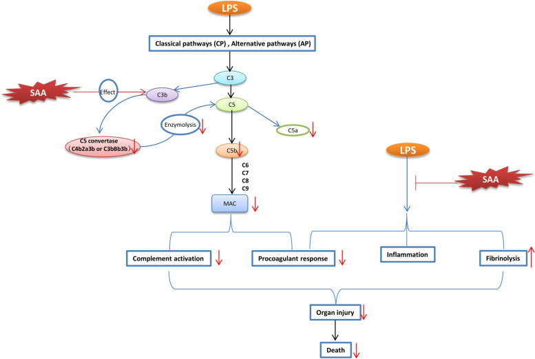
Introduction: Disseminated intravascular coagulation (DIC) is a syndrome characterized by coagulopathy, microthrombus, and multiple organ failure. The complement system in DIC is overactivated, and the functions of complement and coagulation pathways are closely related. Our previous screening revealed that salvianolic acid A (SAA) has anti-complement activity. The hyper-activated complement system was involved in the lipopolysaccharide (LPS) induced DIC in rats. The effects of SAA anti-complement action on LPS-induced DIC in rats were investigated.

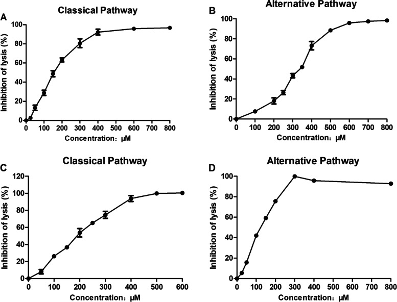
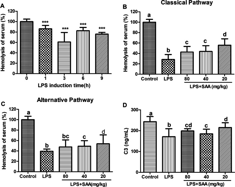
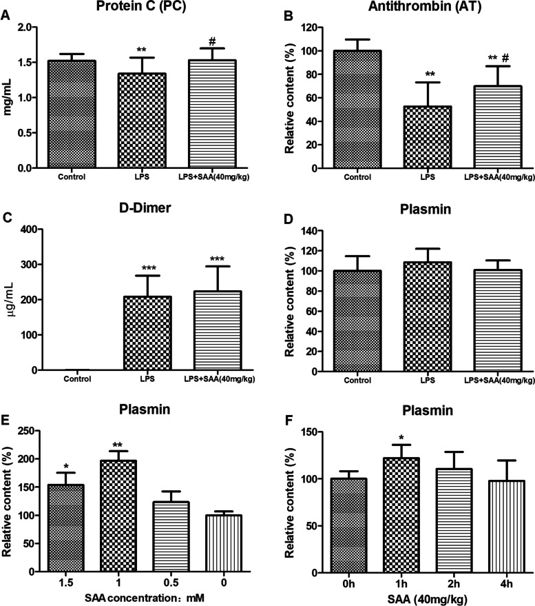
Methods: The complement activity of the classical pathway and alternative pathway was detected through an in vitro hemolysis assay. The binding sites of SAA and complement C3b were predicted by molecular docking. LPS-induced disseminated coagulation experiments were performed on male Wistar rats to assess coagulation function, complement activity, inflammation, biochemistry, blood routine, fibrinolysis, and survival.

Results: SAA had an anti-complement activity in vivo and in vitro and inhibited the complement activation in the classical and alternative pathway of complement. The infusion of LPS into the rats impaired the coagulation function, increased the plasma inflammatory cytokine level, complemented activation, reduced the clotting factor levels, fibrinogen, and platelets, damaged renal, liver, and lung functions, and led to a high mortality rate (85%). SAA treatment of rats inhibited complement activation and attenuated the significant increase in D-dimer, interleukin-6, alanine aminotransferase, and creatinine. It ameliorated the decrease in plasma levels of fibrinogen and platelets and reversed the decline in activity of protein C and antithrombin III. The treatment reduced kidney, liver, and lung damage, and significantly improved the survival rate of rats (46.2 and 78.6% for the low- and high-dose groups, respectively).

Conclusion: SAA reduced LPS-induced DIC by inhibiting complement activation. It has considerable potential in DIC treatment.

Keywords: C3b; Complement system; Disseminated intravascular coagulation; Lipopolysaccharide; Salvianolic acid A.


Conflict of interest statement

The authors declare that they have no conflicts of interest regarding this work.

Figures

Fig.1
Fig.1
In vitro anti-complement activity of SAA. Inhibition of SAA on the classical pathway (A and C) and alternative pathway (B and D) of the complement system. A and B are the rat serum; C and D are the NHS. Results are expressed as percent inhibition of hemolysis. Data were presented as mean ± SD (n = 3)
Fig.2
Fig.2
Prediction of the targets of SAA. A Effect of SAA on the formation of C3/C5 convertase of the complement classical pathway. Different concentrations of SAA were incubated with rat serum and sheep red blood cells for 3 min; after the sheep red blood cells were washed, rat serum containing 0.04 mol/L EDTA was added to initiate hemolysis. The absorbance was measured at 412 nm. Data were presented as mean ± SD (n = 4). B Contents of complement component C3 in the supernatant of classical pathway hemolysis experiment. Different concentrations of SAA were incubated with rat serum and sheep red blood cells for 30 min, centrifuge was performed to obtain the supernatant, and ELISA detection of the C3 content was performed. Data were presented as mean ± SD (n = 3). ***P < 0.001 compared with the serum group, statistical tests used were t-tests. C Map of 10 conjugation sites of SAA and C3b protein. D Conjugation diagram of SAA and C3b protein site 1
Fig.3
Fig.3
Effect of SAA on complement activity in LPS-induced rats. A Hemolytic activity of the serum at LPS induced at different times. Data were listed as mean ± SD (n = 4), ***P < 0.001 compared with the 0 h group, statistical tests used were t-test. The hemolytic activity of CP (B) and AP (C) of the serum was obtained at 3 h. D Contents of complement component C3 in the serum were collected at 3 h. Data were presented as mean ± SD (n = 5), statistical analysis used were ANOVA (P<0.05), letters marked between different groups, if one is the same, the difference is not significant; if they are all different, the difference is significant
Fig.4
Fig.4
Protective effect of SAA on LPS-induced DIC rats. DIC was induced by LPS. A total of 20 and 40 mg/kg SAA was administered by i.p. injection 1 h after, and 15 mg/kg LPS was administer by intravenous infusion. Survival was monitored for over 168 h. *P < 0.05 compared with the LPS group, statistical tests used were t-test
Fig.5
Fig.5
Parameters of SAA on the coagulation in rats with LPS-induced DIC. A Protein C contents in plasma. B Antithrombin contents in plasma. C D-Dimer contents in plasma. D Plasmin contents in plasma. E SAA and rat whole blood were bathed at 37 °C and centrifuged to obtain the supernatant, and the chromogenic substrate S2251 was used to detect plasmin contents. F A total of 40 mg/kg SAA was injected intraperitoneally, and the chromogenic substrate S2251 was used to detect plasma plasmin contents. Data are presented as the mean ± SD. A, B, C, and D, n = 10, **P < 0.01, and ***P < 0.001 compared with the Control group, #P < 0.05 compared with the LPS group. E n = 5, *P < 0.05, and **P < 0.01 compared with the 0 group. F n = 5, *P < 0.05, compared with the 0 h group. Statistical tests used were t-tests
Fig.6
Fig.6
Effects of SAA on the complement activity in LPS-induced DIC. Hemolytic activity of classical pathway (A) and alternative pathway (B) of the serum. C Contents of complement component C3 in serum. D Hemolytic activity of the serum under SAA inducement at different times. Data were presented as the mean ± SD. A, B, and C, n = 10, *P < 0.05, **P < 0.01, and ***P < 0.001 compared with the Control group, #P < 0.05, and ##P < 0.01 compared with the LPS group. D n = 5, *P < 0.05, and ***P < 0.001 compared with the 0 h group. Statistical tests used were t-tests
Fig.7
Fig.7
Effects of SAA on inflammatory cytokine levels in LPS-induced DIC. At 3 h after LPS administration, (A) TNF-α, (B) IL-1β, (C) IL-6, and (D) IL-8 contents in the serum were detected. Data were presented as the mean ± SD. n = 9 in the control group, n = 11 in the LPS group, and n = 10 in the LPS + SAA (40 mg/kg) group. *P < 0.05, **P < 0.01, and ***P < 0.001compared with the Control group. #P < 0.05 compared with the LPS group. Statistical tests used were t-tests
Fig.8
Fig.8
Protective effects of SAA on the organ injury and function in LPS-induced DIC rats. At 3 h after LPS administration, an automatic analyzer was used to detect the ALT (A) and Cre (B) levels in plasma. Data were presented as the mean ± SD. n = 8 in the control group, n = 11 in the LPS group, and n = 9 in the LPS + SAA (40 mg/kg) group. **P < 0.01, ***P < 0.001 compared with the Control group. #P < 0.05, ###P < 0.001 compared with the LPS group. ALT, alanine aminotransferase. Cre, creatinine. At 3 h after LPS administration, lung histopathology of rats (H&E staining): lung H&E staining (C) and lung injury score (D). The original magnifications are 100× and 400×. The red, green, blue, yellow, and orange arrows indicate alveolar epithelial cells, lymphocytes, neutrophils, alveolar macrophages, and fibroblasts, respectively. Results are presented as mean ± SD (n = 6 in each group). ***P < 0.001 compared with the Control group. ###P < 0.001 compared with the LPS group. Statistical tests used were t-tests
Fig. 9
Fig. 9
Schematic drawing of pharmacological effects and mechanisms of SAA

References

    1. Thachil J. Disseminated intravascular coagulation - new pathophysiological concepts and impact on management. Expert Rev Hematol. 2016;9(8):803–814. doi: 10.1080/17474086.2016.1203250. - DOI - PubMed
    1. Levi M, Sivapalaratnam S. Disseminated intravascular coagulation: an update on pathogenesis and diagnosis. Expert Rev Hematol. 2018;11(8):663–672. doi: 10.1080/17474086.2018.1500173. - DOI - PubMed
    1. Iba T, Levi M, Levy JH. Sepsis-induced coagulopathy and disseminated intravascular coagulation. Semin Thromb Hemost. 2020;46(1):89–95. doi: 10.1055/s-0039-1694995. - DOI - PubMed
    1. Hayakawa M. Pathophysiology of trauma-induced coagulopathy: disseminated intravascular coagulation with the fibrinolytic phenotype. J Intensive Care. 2017;5:14. doi: 10.1186/s40560-016-0200-1. - DOI - PMC - PubMed
    1. Levi M. Management of cancer-associated disseminated intravascular coagulation. Thromb Res. 2016;140(Suppl 1):S66–70. doi: 10.1016/S0049-3848(16)30101-3. - DOI - PubMed

LinkOut - more resources