

Ameloblastoma: An Updated Narrative Review of an Enigmatic Tumor
- PMID: 36127985
- PMCID: PMC9481193
- DOI: 10.7759/cureus.27734
Ameloblastoma: An Updated Narrative Review of an Enigmatic Tumor
Abstract
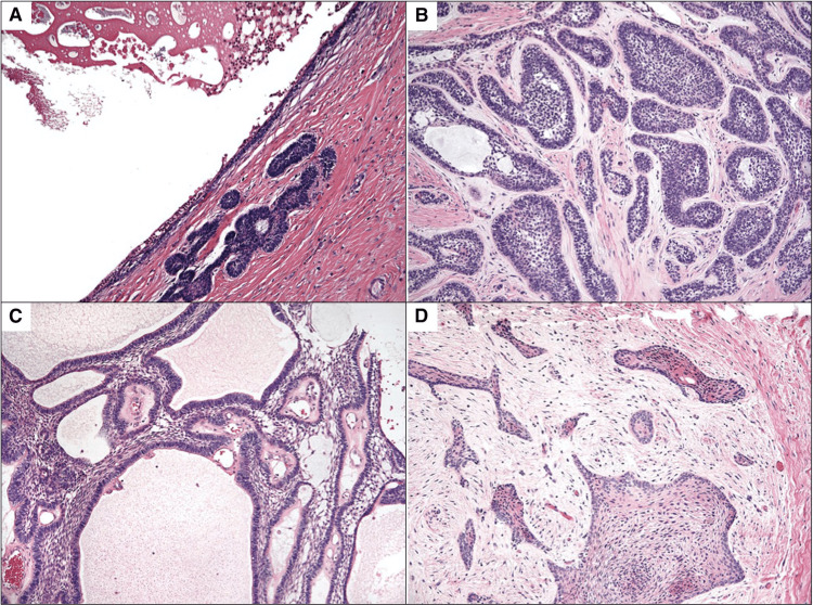
Ameloblastoma is one of the most common benign odontogenic tumors of the jaw that constitutes about 10% of all tumors that arise in the mandible and maxilla. It is a slow-growing but locally invasive tumor that presents with painless swelling of the mandible or maxilla. The World Health Organization (WHO) classification of 2017 describes ameloblastomas of the following four types: ameloblastoma; unicystic ameloblastoma; extraosseous/peripheral ameloblastoma; and metastasizing ameloblastoma. The diagnosis of ameloblastoma requires computerized tomography (CT) imaging as well as a biopsy. A biopsy is helpful in differentiating ameloblastoma from ossifying fibroma, osteomyelitis, giant cell tumor, cystic fibrous dysplasia, myeloma, and sarcoma. The best treatment of ameloblastoma is aggressive en bloc resection with simultaneous reconstruction. The high recurrence rate and large tissue defects have been long-standing issues in the treatment of ameloblastoma. Recent molecular developments strongly suggest the possibility of targeted therapy with better outcomes in ameloblastomas. We present a detailed updated narrative review of our current understanding and management of this enigmatic tumor.
Keywords: adamantinoma; ameloblastoma; brafv600e mutation; jaw neoplasms; mandible; mandibular neoplasms; maxilla; maxillary neoplasms; multicystic ameloblastoma; odontogenic tumors.
Copyright © 2022, Ghai et al.
Conflict of interest statement
The authors have declared that no competing interests exist.
Figures
Similar articles
-
Unicystic ameloblastoma: a clinicopathologic study of 33 Chinese patients.Am J Surg Pathol. 2000 Oct;24(10):1385-92. doi: 10.1097/00000478-200010000-00008. Am J Surg Pathol. 2000. PMID: 11023100
-
An Unusual Case Report of Unicystic Ameloblastoma of the Mandible.J Pharm Bioallied Sci. 2024 Feb;16(Suppl 1):S955-S959. doi: 10.4103/jpbs.jpbs_568_23. Epub 2024 Feb 29. J Pharm Bioallied Sci. 2024. PMID: 38595394 Free PMC article.
-
Assessment of BRAFV600E and SMOF412E mutations in epithelial odontogenic tumours.Tumour Biol. 2015 Jul;36(7):5649-53. doi: 10.1007/s13277-015-3238-0. Epub 2015 Feb 18. Tumour Biol. 2015. PMID: 25854168
-
Ameloblastoma: biological profile of 3677 cases.Eur J Cancer B Oral Oncol. 1995 Mar;31B(2):86-99. doi: 10.1016/0964-1955(94)00037-5. Eur J Cancer B Oral Oncol. 1995. PMID: 7633291 Review.
-
Is there a role for enucleation in the management of ameloblastoma?Int J Oral Maxillofac Surg. 2009 Aug;38(8):807-12. doi: 10.1016/j.ijom.2009.02.018. Epub 2009 Mar 17. Int J Oral Maxillofac Surg. 2009. PMID: 19297131 Review.
Cited by
-
Frequency of BRAF V600E immunoexpression in ameloblastomas: a multi-institutional analysis of 86 cases in Latin America and comprehensive review of the literature.Med Oral Patol Oral Cir Bucal. 2024 Jul 1;29(4):e509-e516. doi: 10.4317/medoral.26493. Med Oral Patol Oral Cir Bucal. 2024. PMID: 38615253 Free PMC article. Review.
-
Factors related to risk of recurrence and recurrence free survival in ameloblastoma of the Jaws: A single centre retrospective analysis.Oral Maxillofac Surg. 2024 Dec 30;29(1):22. doi: 10.1007/s10006-024-01321-3. Oral Maxillofac Surg. 2024. PMID: 39739058
-
A Retrospective Analysis of 129 Ameloblastoma Cases: Clinical and Demographical Trends from a Single Institution.J Racial Ethn Health Disparities. 2025 Jun;12(3):1612-1620. doi: 10.1007/s40615-024-01993-3. Epub 2024 Apr 12. J Racial Ethn Health Disparities. 2025. PMID: 38607614 Free PMC article.
-
Desmoplastic histological subtype of ameloblastoma in 16 dogs.Front Vet Sci. 2024 Apr 4;11:1362237. doi: 10.3389/fvets.2024.1362237. eCollection 2024. Front Vet Sci. 2024. PMID: 38638641 Free PMC article.
-
NKG2D (Natural Killer Group 2, Member D) ligand expression and ameloblastoma recurrence: a retrospective immunohistological pilot study.BMC Oral Health. 2024 Sep 17;24(1):1102. doi: 10.1186/s12903-024-04873-8. BMC Oral Health. 2024. PMID: 39289711 Free PMC article.
References
-
- Ameloblastoma. Masthan KM, Anitha N, Krupaa J, Manikkam S. http://www.jpbsonline.org/article.asp?issn=0975-7406;year=2015;volume=7;... J Pharm Bioallied Sci. 2015;7:0–70. - PMC - PubMed
-
- Report of the amputations of the lower jaw. Cusack J. Dublin Hosp Rec. 1827;4:1–38.
-
- Sur le role des debris epitheliaux papdentaires. Malassez L. Arch Physiol Norm Pathol. 1885;5:309–340.
-
- The need of a standardized surgical and pathological classification of tumors and anomalies of dental origin. Ivey R, Churchill H. Am Assoc Dent Sch Trans. 1930;7:240–245.
-
- Vered M, Muller S, Heikinheimo K. IARC WHO Classification of Tumours. 4th Edition, Volume 9. Vol. 215. Lyon, France: IARC Press; 2017. Ameloblastoma; p. 8.
Publication types
LinkOut - more resources
Full Text Sources
Research Materials