Case Reports
. 2022 Aug;13(8):402-407.
doi: 10.14740/jmc3952. Epub 2022 Aug 19.

Uterine Cervical Adenosarcoma Showing an Endophytic Growth Pattern

Affiliations
Case Reports

Uterine Cervical Adenosarcoma Showing an Endophytic Growth Pattern

Eri Lin-Satoi et al. J Med Cases. 2022 Aug.

Abstract

Adenosarcomas are biphasic neoplasms that usually originate in the uterine corpus and comprise a benign epithelial component and a malignant stromal component. Uterine adenosarcomas typically present with abnormal genital bleeding, an enlarged uterus, and a tumor that protrudes into the endometrial cavity. These tumors rarely protrude through the cervical os and are often misdiagnosed as cervical polyps. We present the case of a patient with cervical adenosarcoma with characteristics different from those reported in previous cases. This tumor showed endophytic growth, which is rare in cervical adenosarcomas. No watery discharge or obvious genital bleeding was noted. Although the tumor measured 4 cm, vaginal bleeding was noted only once at 6 months before diagnosis and was in the form of faint brown discharge.

Keywords: Adenosarcoma; Endophytic growth; Uterine cervix.


Conflict of interest statement

None to declare.

Figures

Figure 1
Figure 1
Fluorodeoxyglucose positron emission tomography-computed tomography indicating increased uptake in the cervix (arrow: cervical tumor).
Figure 2
Figure 2
(a) Diffusion-weighted image showing a heterogeneous high-intensity tumor (arrow). (b) Apparent diffusion coefficient image showing a tumor with mild diffusion restriction (arrow).
Figure 3
Figure 3
Multiple hyperintense small cysts inside the tumor on T2-weighted imaging (arrow).
Figure 4
Figure 4
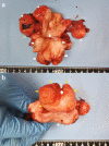
(a) Gross pathological specimen showing a mass occupying the entire cervix (arrowhead: ovary, star: leiomyoma, arrow: cervical tumor). (b) Cervix replaced by the tumor, as seen from the vaginal side (arrow: external os with an incision at 12 o’clock; yellow arrow: cervix; arrowhead: vaginal wall).
Figure 5
Figure 5
(a) Low-power field showing characteristic leaf-like architecture (hematoxylin and eosin staining, magnification × 10). (b) Papillary stromal fronds lined by benign epithelium accompanied with squamous metaplasia. The stromal cells show mild nucleal atypia and mild pleomorphism (hematoxylin and eosin staining, magnification × 200).
Figure 6
Figure 6
(a) Subepithelial condensation of the stroma; periglandular cuffing (hematoxylin and eosin staining, magnification × 40). (b) Hypercellular stroma and high cellularity beneath the epithelium (hematoxylin and eosin staining, magnification × 100).
Figure 7
Figure 7
Stromal atypia with mitosis in the stromal element (arrow) (hematoxylin and eosin staining, magnification × 400).
Figure 8
Figure 8
Immunohistochemical findings of the tumor. (a) Stromal cell cytoplasm positive for CD10 (magnification, ×100); (b) glandular epithelia nuclei positive but stromal cell nuclei negative for estrogen receptors (magnification, ×100); (c) both glandular epithelia nuclei and stromal cell nuclei negative for progesterone receptors (magnification, ×100); (d) stromal cell showing wild-type pattern for P53 (magnification, ×40).

References

    1. D'Angelo E, Prat J. Uterine sarcomas: a review. Gynecol Oncol. 2010;116(1):131–139. doi: 10.1016/j.ygyno.2009.09.023. - DOI - PubMed
    1. Harlow BL, Weiss NS, Lofton S. The epidemiology of sarcomas of the uterus. J Natl Cancer Inst. 1986;76(3):399–402. - PubMed
    1. Abeler VM, Royne O, Thoresen S, Danielsen HE, Nesland JM, Kristensen GB. Uterine sarcomas in Norway. A histopathological and prognostic survey of a total population from 1970 to 2000 including 419 patients. Histopathology. 2009;54(3):355–364. doi: 10.1111/j.1365-2559.2009.03231.x. - DOI - PubMed
    1. Verschraegen CF, Vasuratna A, Edwards C, Freedman R, Kudelka AP, Tornos C, Kavanagh JJ. Clinicopathologic analysis of mullerian adenosarcoma: the M.D. Anderson Cancer Center experience. Oncol Rep. 1998;5(4):939–944. doi: 10.3892/or.5.4.939. - DOI - PubMed
    1. Togami S, Kawamura T, Fukuda M, Yanazume S, Kamio M, Kobayashi H. Clinical management of uterine cervical mullerian adenosarcoma: A clinicopathological study of six cases and review of the literature. Taiwan J Obstet Gynecol. 2018;57(4):479–482. doi: 10.1016/j.tjog.2018.04.032. - DOI - PubMed

Publication types

LinkOut - more resources