Prediction of the functional and radiological outcome on the basis of independent factors with special emphasis on the use of 3D printed models in craniovertebral junction surgery
- PMID: 36128135
- PMCID: PMC9479533
- DOI: 10.25259/SNI_998_2021
Prediction of the functional and radiological outcome on the basis of independent factors with special emphasis on the use of 3D printed models in craniovertebral junction surgery
Abstract
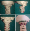
Background: The aim of the study was to evaluate the advantage of performing planned surgery using customized three-dimensional (3D) printed models versus performing surgery without using 3D printed models in patients with craniovertebral junction (CVJ) anomalies and traumatic CVJ fractures and dislocations.
Methods: Forty-two patients with CVJ anomalies, who were planned for operative intervention in the Department of Neurosurgery at SMS Hospital from March 2019 to February 2021, were randomly divided into two groups and analyzed. First group was operated after rehearsal on a customized 3D printed model whereas the second group underwent operative intervention without the rehearsal of surgery on the 3D printed model.
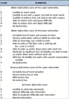
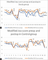
Results: Forty-two patients were enrolled for the study. Twenty-five of these patients had developmental CVJ anomalies, 16 had post traumatic Atlantoaxial dislocation (AAD), and one had congenital AAD. Twenty-three patients underwent surgical intervention using 3D printed models and 19 without using 3D printed models. The outcome in the two groups was compared using modified Japanese orthopedic association score (mJOA), recovery rate, incidence of complications such as screw malposition, postoperative neurological deterioration, vertebral artery (VA) injury, and radiological improvement based on Atlanto-Dental interval, the distance of the tip of dens from Wackhenheims clivus canal line, and the distance of tip of dens from the Chamberlain's line. The improvement in mJOA score postoperatively was found to be statistically significant in study group (P < 0.001) as compared to control group (P = 0.06). Recovery rate was better in study group than in control group (P = 0.023). In study group, the incidence of screw malposition and VA injury was lower than control group. Three patients deteriorated neurologically postoperatively in the control group and none in the study group. The average improvements in the radiological parameters were found to be better in study group as compared to control group postoperatively.
Conclusion: The authors conclude that 3D printed models are extremely helpful in analyzing joints and VA anatomy preoperatively and are helpful in unmasking any abnormal bony and vascular anatomy effectively, making the surgeon confident about the placement of the screws intraoperatively. These 3D models help in intraoperative error minimization with better neurological outcomes in postoperative period. In our opinion, these models should be included as a basic investigation tool in patients of CVJ abnormalities. The models also offer other advantages such as preoperative simulation, teaching modules, and patient education.
Keywords: 3D printed model; Atlantoaxial dislocation; Basilar invagination; C1–C2; Craniovertebral junction abnormality; Occiput–C2.
Copyright: © 2022 Surgical Neurology International.
Conflict of interest statement
There are no conflicts of interest.
Figures


References
-
- Abumi K, Shono Y, Ito M, Taneichi H, Kotani Y, Kaneda K. Complications of pedicle screw fixation in reconstructive surgery of the cervical spine. Spine (Phila Pa 1976) 2000;25:962–9. - PubMed
-
- Behari S, Kiran Kumar MV, Banerji D, Chhabra DK, Jain VK. Atlantoaxial dislocation associated with the maldevelopment of the posterior neural arch of axis causing compressive myelopathy. Neurol India. 2004;52:489–91. - PubMed
LinkOut - more resources
Full Text Sources
Miscellaneous
