. 2022 Aug 18:12:49.
doi: 10.25259/JCIS_76_2022. eCollection 2022.

Catheter-directed computed tomography angiography: A pictorial essay

Affiliations

Catheter-directed computed tomography angiography: A pictorial essay

Abheek Ghosh et al. J Clin Imaging Sci. .

Abstract

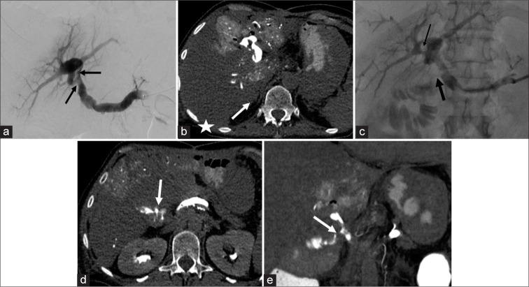
Catheter-directed computed tomography angiography (CDCTA) is an imaging technique where CT images are acquired after selective catheterization of a vessel. Images obtained in this fashion provide several advantages over conventional imaging techniques such as fluoroscopic angiography, digital subtraction angiography, cone-beam CT, and conventional CT angiography. At this point, there is still limited literature on the subject, with prior studies examining a small number of potential uses. The goal of this pictorial essay is to illustrate our single tertiary care center experience using CDCTA.

Keywords: Catheter-directed computed tomography angiography; Computed tomography angiography; Interventional radiology.


Conflict of interest statement

There are no conflicts of interest.

Figures

Figure 1:
Figure 1:
(a) 72-year-old male with HCC, and history of bilaterhal TACE and radiation segmentectomy presents with residual disease. Plan for vessel mapping prior to Y-90 radioembolization. Case above provides an example where CBCT from the left hepatic artery is unable to fully characterize the treatment region (arrow) secondary to an inadequate field of view as well as poor spatial resolution. (b) Left hepatic artery sub-selective CDCTA reveals an area of nodular enhancement (arrow) clearly separate from the background peri-treatment change, axial view. (c) Coronal view complimentary to Figure 1b. (d) In contrast, axial and coronal CDCTAs of another previously treated lesion from the right hepatic artery only show post-treatment changes without evidence of tumor recurrence (arrow). Increased contrast conspicuity and spatial resolution of CDCTA allowed for this delineation. (e) Coronal view complimentary to Figure 1d.
Figure 2:
Figure 2:
(a) 46-year-old female with ovarian fibroids and menorrhagia. Plan for uterine fibroid embolization (UFE). Mapping for UFE can be complicated by distorted pelvic anatomy (i.e. enlarged uterus) and the possibility of collateral uterine supply. Initial abdominal aortic DSA above demonstrates occlusion of the left internal iliac artery at its origin (white arrow), and no significant right uterine artery (black arrow). (b) The ovarian vessels were presumed to be the dominant blood supply; however, aortic fluoroscopic angiograpy only faintly revealed the left ovarian artery (arrow). (c) Aortic CDCTA revealed the origin of both ovarian vessels (white arrows). (d) Aortic CDCTA also confirmed a tortuous hypertrophied course into the pelvis to supply the uterus (white arrows). (e) After successful right ovarian artery cannulation, DSA revealed prominent uterine uptake (arrow).
Figure 3:
Figure 3:
(a) 19-year-old female s/p MVC with rib fractures and bloody output from chest tube. Plan for angiogram with possible subsequent embolization. In cases of intercostal artery hemorrhage, identifying the involved thoracic level is important for quick cannulation and embolization. Conventional axial CTA did not demonstrate active arterial extravasation, with only non-localized pooling of contrast in the pleural space in the venous phase (arrow). (b) CDCTA via the aorta identified active extravasation in the 7th intercostal artery (arrow), axial view. (c) Coronal view complimentary to Figure 3b. (d) CDCTA via the aorta also revealed active extravasation of the 8th intercostal artery (arrow), axial view. (e) Coronal view complimentary to Figure 3d. (f) Targeted cannulation and arteriogram confirmed the findings. Increased contrast conspicuity over conventional CTA allowed for visualization of the bleed (arrow).
Figure 4:
Figure 4:
(a) 67-year-old female with history of colonic diverticulosis presents with lower GI bleeding, and decreased hemoglobin level. Detecting GI hemorrhage with conventional imaging methods is limited due to the intermittent nature of active arterial extravasation as well as the sensitivity of the exam. Axial conventional CTA demonstrated no extravasated contrast. (b) Tagged red blood cell nuclear machine failed to identify the bleed in this patient with clinically significant hemorrhage. (c) During a period of suspected ongoing hemorrhage, DSA via the SMA identified a region of possible extravasation from a right colic branch (arrow). (d) Sub-selective CDCTA demonstrated active extravasation from the suspected right colic branch (arrow), axial view. (e) Coronal view complimentary to Figure 4d. (f) Venous CDCT further confirmed the findings with pooling of extravasated blood.
Figure 5:
Figure 5:
(a) 65-year-old female with suspected infected portal venous clot. Plan for portal venogram and suction thrombectomy. Removal of a portal venous clot unmasked a portal venous-bowel fistula in a patient with a complex abdominal surgical history, including recent Whipple for pancreatic carcinoma. Initial trans-splenic portal venogram demonstrates scattered filling defects (black arrows). (b) Portal venous CDCT demonstrates the clot (arrow) as well as non-opacification of the right portal venous system (star) due to the chronic occlusive right portal vein thrombus. (c) After partial suction thrombectomy, a portal vein-bowel fistula was revealed on portal venogram (black arrows). (d) Portal venous CDCT more precisely described the fistula as involving the choledochojejunostomy loop used in the patient’s Whipple (arrow), axial view. High resolution CDCT in a large field of view aided in surgical planning in the setting of prior abdominal surgeries. (e) Coronal view complimentary to Figure 5d.

Similar articles

References

    1. Isaacson AJ, Burke LM, Vallabhaneni R, Farber MA. Ultralow iodine dose transarterial catheter-directed CT angiography for fenestrated endovascular aortic repair planning. Ann Vasc Surg. 2016;35:234–7. doi: 10.1016/j.avsg.2016.01.045. - DOI - PubMed
    1. Özgen A, Sanioglu S, Bingöl UA. Intra-arterial ultra-low-dose CT angiography of lower extremity in diabetic patients. Cardiovasc Interv Radiol. 2016;39:1165–9. doi: 10.1007/s00270-016-1358-6. - DOI - PubMed
    1. Goh WX, Leong S, Too CW, Cheng LT, Saffaru SE, Lee RZ, et al. Catheter-directed computed tomography hepatic angiography for Yttrium-90 selective internal radiotherapy of hepatocellular carcinoma reduces prophylactic embolization of extrahepatic vessels. Cardiovasc Interv Radiol. 2019;43:478–87. doi: 10.1007/s00270-019-02362-y. - DOI - PubMed

LinkOut - more resources