

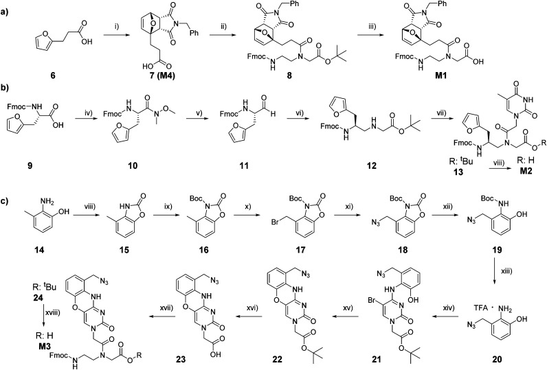
Synthesis and structure-activity relationship of peptide nucleic acid probes with improved interstrand-crosslinking abilities: application to biotin-mediated RNA-pulldown
- PMID: 36128507
- PMCID: PMC9428673
- DOI: 10.1039/d2cb00095d
Synthesis and structure-activity relationship of peptide nucleic acid probes with improved interstrand-crosslinking abilities: application to biotin-mediated RNA-pulldown
Abstract
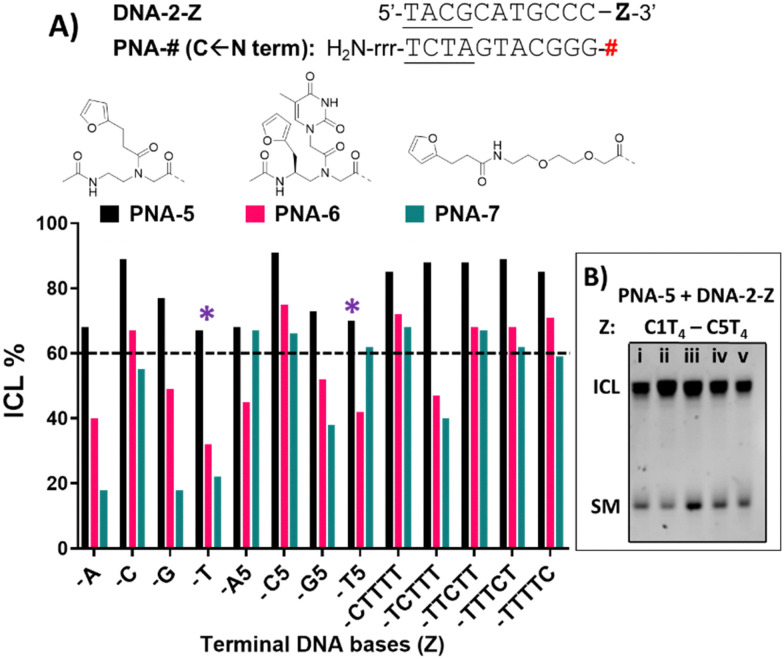
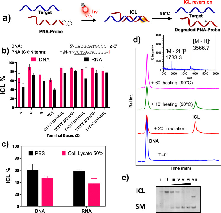
The development of interstrand-crosslinking (ICL) probes for the covalent targeting of DNA and RNA sequences of interest has been extensively reported in the past decade. However, most of the reactions reported so far induce the formation of a stable adduct that cannot be reverted, thus rendering these chemistries less useful in applications where the reversibility of the reaction is needed for further downstream processing of the targeted and isolated sequences, such as enzymatic amplification steps. In this work, we report on the reversibility of the furan-mediated ICL reaction. ICL formation can be conveniently triggered by either chemical (N-bromo succinimide, NBS) or luminous stimuli (visible light irradiation in presence of a photosensitizer) and quantitative reversion can be achieved by heating the crosslinked sample at 95 °C, while maintaining the structure of the DNA/RNA targets intact. As a proof-of-concept and showing the benefits of the ICL reversibility, we apply furan-mediated ICL to the pulldown of a target RNA strand from cell lysate.
This journal is © The Royal Society of Chemistry.
Conflict of interest statement
There are no conflicts to declare.
Figures


Similar articles
-
Furan-based (photo)oxidation reactions and their application in nucleic acid and protein targeting.Methods. 2023 Oct;218:189-197. doi: 10.1016/j.ymeth.2023.08.011. Epub 2023 Aug 18. Methods. 2023. PMID: 37597698
-
Pyrrolidinyl Peptide Nucleic Acid Probes Capable of Crosslinking with DNA: Effects of Terminal and Internal Modifications on Crosslink Efficiency.Chembiochem. 2021 Jan 5;22(1):241-252. doi: 10.1002/cbic.202000589. Epub 2020 Oct 23. Chembiochem. 2021. PMID: 32889765
-
Furan-modified PNA probes for covalent targeting and ligation of nucleic acids.Methods. 2023 Oct;218:210-223. doi: 10.1016/j.ymeth.2023.08.010. Epub 2023 Aug 19. Methods. 2023. PMID: 37604247
-
Coordinated Cut and Bypass: Replication of Interstrand Crosslink-Containing DNA.Front Cell Dev Biol. 2021 Jun 28;9:699966. doi: 10.3389/fcell.2021.699966. eCollection 2021. Front Cell Dev Biol. 2021. PMID: 34262911 Free PMC article. Review.
-
Mechanisms of interstrand DNA crosslink repair and human disorders.Genes Environ. 2016 May 1;38:9. doi: 10.1186/s41021-016-0037-9. eCollection 2016. Genes Environ. 2016. PMID: 27350828 Free PMC article. Review.
Cited by
-
A red light-triggered chemical tool for sequence-specific alkylation of G-quadruplex and I-motif DNA.Nucleic Acids Res. 2023 May 22;51(9):4112-4125. doi: 10.1093/nar/gkad189. Nucleic Acids Res. 2023. PMID: 36971129 Free PMC article.
-
Impact of charges on the hybridization kinetics and thermal stability of PNA duplexes.Org Biomol Chem. 2024 Jul 17;22(28):5759-5767. doi: 10.1039/d4ob00887a. Org Biomol Chem. 2024. PMID: 38920402 Free PMC article.
References
-
- Seeman N. C. Sleiman H. F. Nat. Rev. Mater. 2018;3:17068. doi: 10.1038/natrevmats.2017.68. - DOI
LinkOut - more resources
Full Text Sources
Research Materials
Miscellaneous