. 2022 Nov;61(5):133.
doi: 10.3892/ijo.2022.5423. Epub 2022 Sep 21.

Isolation and analysis of tumor‑derived extracellular vesicles from head and neck squamous cell carcinoma plasma by galectin‑based glycan recognition particles

Affiliations

Isolation and analysis of tumor‑derived extracellular vesicles from head and neck squamous cell carcinoma plasma by galectin‑based glycan recognition particles

Laura Benecke et al. Int J Oncol. 2022 Nov.

Abstract

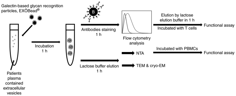
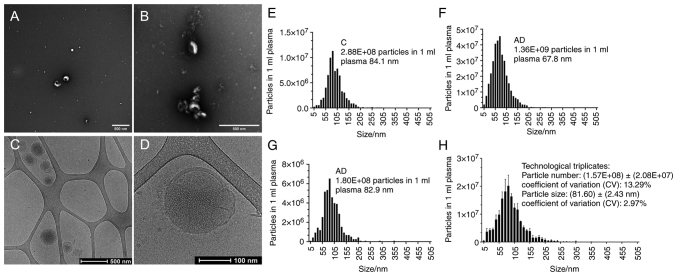
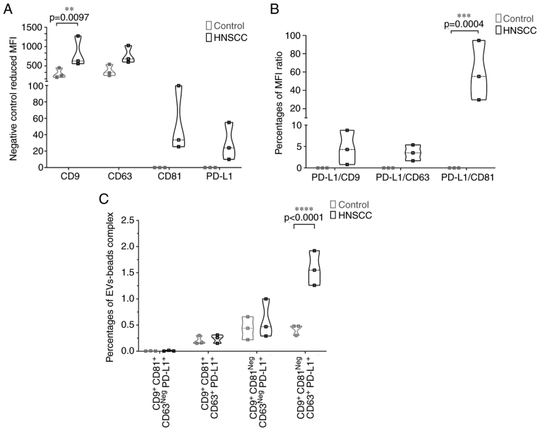
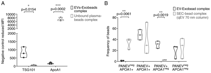
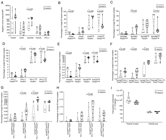
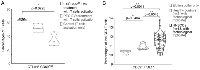
Extracellular vesicles (EVs) have recently come into the spotlight as potential cancer biomarkers. Isolation of pure EVs is complex, so wider use requires reliable and time‑efficient isolation methods. In the present study, galectin‑based magnetic glycan recognition particles, EXÖBead® were investigated for their practicality as a novel EV isolation technique, exemplified here for squamous cell carcinoma of the head and neck. Analysis of the isolation method showed a high concentration of pure EVs with detection of specific EV markers such as CD9, CD63, CD81 and TSG101. No apolipoprotein A1 was shown in the isolates, indicating low contamination of this isolation technique compared with size exclusion chromatography. In addition, common leukocyte antigen (CD45), three HNSCC [epithelial cell adhesion molecule (EpCAM), pan‑cytokeratin and programmed death‑ligand 1 (PD‑L1)] and PanEV markers (premixed CD9, CD63 and CD81 antibodies) were measured by bead‑based flow cytometry (BFC). BFC revealed that CD45Neg PanEV+, EpCAM+ PanEV+ and PD‑L1+ PanEV+ were significantly higher in tumor patients compared with healthy control plasma. CD45Neg PanEV+ and CD45+ PanEV+ carrying two or three HNSCC biomarkers were also significantly higher in tumor patients compared with healthy controls (BFC). Comparison of the functional immunosuppression effect of eluted tumor patient plasma EVs from EXÖBead® and commercial polyethylene glycol isolation showed a significant tumor‑dependent increase in concentration of EVs. A peripheral blood mononuclear cell activation assay also showed that the T‑cell functionality of tumor patient plasma EVs isolated with EXÖBead® was preserved in vitro. In conclusion, isolation using galectin‑based magnetic glycan recognition particles is a novel method for isolating plasma EVs with low lipoprotein contamination. Bead‑based flow cytometry provided an easy way to understand EV subpopulations. EXÖBead® therefore showed great potential as a new isolation tool with high throughput capacity that could potentially be used in a clinical setting.

Keywords: beads‑based flow cytometry; biomarker; exosomes; extracellular vesicles; galectin‑based glycan recognition particles; head and neck squamous cell carcinoma; novel isolation technique; tumor‑derived extracellular vesicles.


Conflict of interest statement

DC is the founder of Biovesicle, Inc. All experiments were conducted without financial contribution, simply scientific advices were obtained. LM and MWP are scientific advisor of Biovesicle, Inc. and did not receive profit.

Figures

Figure 1
Figure 1
Workflow for extracellular vesicles isolation and analysis by using galectin-based glycan recognition particles, EXÖBead®.
Figure 2
Figure 2
EV morphology and size distribution. (A and B) Eluted EVs by EXÖBead® isolation in transmission electron microscopy. (C and D) Eluted EVs by EXÖBead® isolation in cryo-electron microscopy. (E-G) Particle size distribution of eluted EVs by EXÖBead® isolation from three individual donors, measured with Zetaview®. (H) Particle size distribution of eluted EVs by EXÖBead® isolation from the same donor with three independent experiments, measured with Zetaview®. EV, extracellular vesicle.
Figure 3
Figure 3
Bead-based flow cytometric analysis of EV surface markers. (A) EV surface markers of plasma EV-EXÖBead® complexes (patients: n=3 and healthy controls: n=3) are shown as reduced geometric MFI of CD9, CD63, CD81 and PD-L1 in the negative control. (B) Plot of the ratio of the MFI of PD-L1 to the MFI of CD9, the MFI of PD-L1 to the MFI of CD63, and the MFI of PD-L1 to the MFI of CD81. (C) CD9+ CD81+ CD63Neg PD-L1+, CD9+ CD81+ CD63+ PD-L1+, CD9+ CD81Neg CD63Neg PD-L1+ and PD-L1+ CD9+ CD81Neg CD63+ of plasma EV-EXÖBead® complex were gated with FlowJo™. Significance was calculated by Two-way ANOVA with Šídák's multiple comparisons test. EV, extracellular vesicle; MFI, mean fluorescence intensity.
Figure 4
Figure 4
Bead-based flow cytometry analysis of EV intracellular marker and non-EV marker. (A) Intracellular EVs markers (TSG101) and non-EV markers (ApoA1) of plasma EVs-EXÖBead® complexes and unbound plasma fraction magnetic bead complexes (n=3) are shown as reduced geometric mean fluorescence intensity in the negative control. (B) PanEV+/Neg and ApoA1+/Neg populations of the plasma EVs-EXÖBead® complex and SEC (Izon qEVoriginal 70) plasma EVs-magnetic beads complex are expressed as percentages by gating with FlowJo™. Significance was calculated using an unpaired t-test. EV, extracellular vesicle.
Figure 5
Figure 5
Bead-based flow cytometric analysis of HNSCC biomarkers. (A) EVs surface marker (PanEV), leukocyte common marker (CD45) and HNSCC markers (EpCAM, PanCK and PD-L1) of plasma EVs-EXÖBead® complexes (patients: n=9 and healthy controls: n=9 are shown as reduced geometric mean fluorescence intensity in the negative control. (B) PanEV+/Neg CD45+/Neg. (C) PanEV+/Neg EpCAM+/Neg. (D) PanEV+/Neg PD-L1+/Neg. (E) PanEV+/Neg PanCK+/Neg. (F) CD45neg PanEV+ EpCAM+/Neg and PD-L1+/Neg. (G) CD45neg PanEV+ PD-L1+ EpCAM+/Neg and PanCK+/Neg. (H) CD45+ PanEV+ PD-L1+ EpCAM+/Neg and PanCK+/Neg of single plasma EVs-EXÖBead® complex were evaluated using FlowJo™. Significance was calculated using an unpaired t-test with Welch's correction. (I) Particle number and size from HNSCC (n=9) and control plasma (n=9) are measured by Zetaview®. Significance was calculated using Kolmogorov Smirnov test. EV, extracellular vesicle; HNSCC, head and neck squamous cell carcinoma; PanCK, pan-cytokeratin; EpCAM, epithelial cell adhesion molecule.
Figure 6
Figure 6
EVs functional assay of T cells and PBMCs activation. (A) A total of 30 out of 200 µl eluted patient plasma EVs from EXÖBead® isolation and PEG EVs were treated with CD4+ T cells in anti-CD2/3/28 antibodies activation condition. The Violin plot shows that CTLA4+ CD69Neg T cells emerged only when treated with eluted patient plasma EVs from EXÖBead®, PEG EVs and T cells activation. Significance was calculated by non-parametric Kruskal-Wallis test with Dunn's multiple comparison test. (B) a total of 5×10⁷ particles of eluted patient or control plasma EVs from EXÖBead® isolation were treated with 1×10⁶ PBMCs (ratio: 50:1) under anti-CD2/3/28 antibody activation conditions. Violin plot showed that CD69+ PD-L1+ live CD4+ T cells were derived from treatment with elution buffer alone, from plasma EVs from HNSCC patients (n=13, with technological triplicate) and from EVs from healthy controls (n=3, with technological triplicate). Significance was calculated by Brown-Forsythe and Welch's ANOVA test with Dunnett's T3 multiple comparisons test. EV, extracellular vesicle; PBMC, peripheral blood mononuclear cells.

References

    1. Raposo G, Nijman HW, Stoorvogel W, Liejendekker R, Harding CV, Melief CJ, Geuze HJ. B lymphocytes secrete antigen-presenting vesicles. J Exp Med. 1996;183:1161–1172. doi: 10.1084/jem.183.3.1161. - DOI - PMC - PubMed
    1. Harding C, Heuser J, Stahl P. Receptor-mediated endocytosis of transferrin and recycling of the transferrin receptor in rat reticulocytes. J Cell Biol. 1983;97:329–339. doi: 10.1083/jcb.97.2.329. - DOI - PMC - PubMed
    1. Lopatina T, Favaro E, Danilova L, Fertig EJ, Favorov AV, Kagohara LT, Martone T, Bussolati B, Romagnoli R, Albera R, et al. Extracellular vesicles released by tumor endothelial cells spread immunosuppressive and transforming signals through various recipient cells. Front Cell Dev Biol. 2020;8:698. doi: 10.3389/fcell.2020.00698. - DOI - PMC - PubMed
    1. Xiao C, Song F, Zheng YL, Lv J, Wang QF, Xu N. Exosomes in head and neck squamous cell carcinoma. Front Oncol. 2019;9:894. doi: 10.3389/fonc.2019.00894. - DOI - PMC - PubMed
    1. Bray F, Ferlay J, Soerjomataram I, Siegel RL, Torre LA, Jemal A. Global cancer statistics 2018: GLOBOCAN estimates of incidence and mortality worldwide for 36 cancers in 185 countries. CA Cancer J Clin. 2018;68:394–424. doi: 10.3322/caac.21492. - DOI - PubMed

MeSH terms