

Adaptation of Fibril-Reinforced Poroviscoelastic Properties in Rabbit Collateral Ligaments 8 Weeks After Anterior Cruciate Ligament Transection
- PMID: 36129552
- PMCID: PMC10023629
- DOI: 10.1007/s10439-022-03081-1
Adaptation of Fibril-Reinforced Poroviscoelastic Properties in Rabbit Collateral Ligaments 8 Weeks After Anterior Cruciate Ligament Transection
Abstract
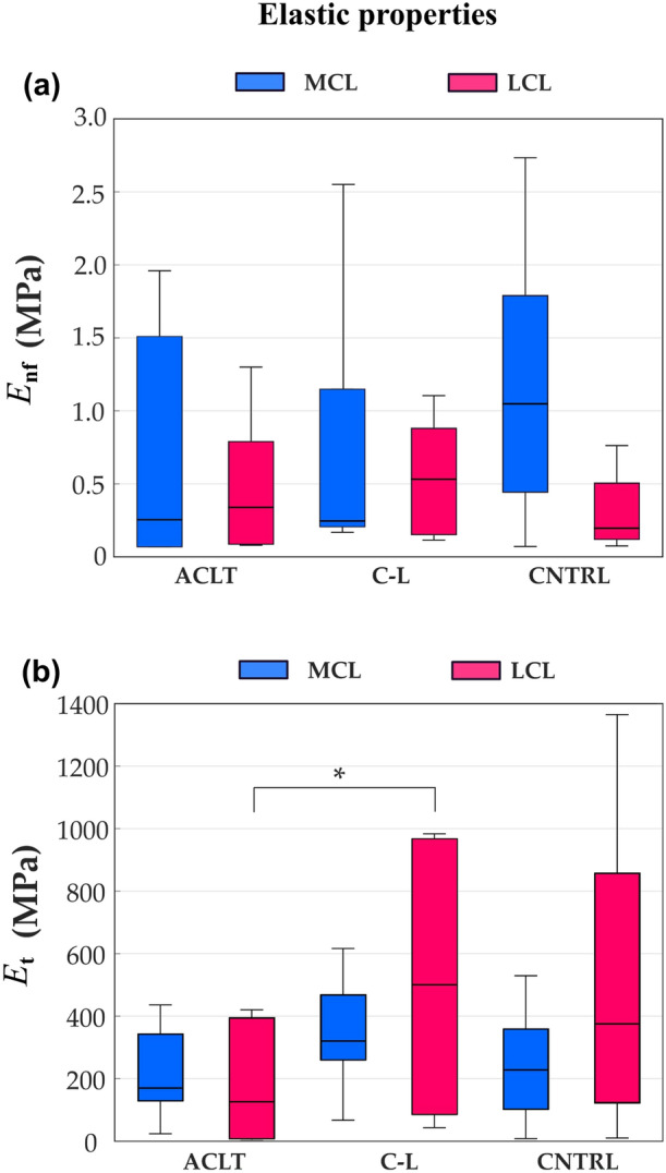
Ligaments of the knee provide stability and prevent excessive motions of the joint. Rupture of the anterior cruciate ligament (ACL), a common sports injury, results in an altered loading environment for other tissues in the joint, likely leading to their mechanical adaptation. In the collateral ligaments, the patterns and mechanisms of biomechanical adaptation following ACL transection (ACLT) remain unknown. We aimed to characterize the adaptation of elastic and viscoelastic properties of the lateral and medial collateral ligaments eight weeks after ACLT. Unilateral ACLT was performed in six rabbits, and collateral ligaments were harvested from transected and contralateral knee joints after eight weeks, and from an intact control group (eight knees from four animals). The cross-sectional areas were measured with micro-computed tomography. Stepwise tensile stress-relaxation testing was conducted up to 6% final strain, and the elastic and viscoelastic properties were characterized with a fibril-reinforced poroviscoelastic material model. We found that the cross-sectional area of the collateral ligaments in the ACL transected knees increased, the nonlinear elastic collagen network modulus of the LCL decreased, and the amount of fast relaxation in the MCL decreased. Our results indicate that rupture of the ACL leads to an early adaptation of the elastic and viscoelastic properties of the collagen fibrillar network in the collateral ligaments. These adaptations may be important to consider when evaluating whole knee joint mechanics after ACL rupture, and the results aid in understanding the consequences of ACL rupture on other tissues.
Keywords: Anterior cruciate ligament transection; Finite element model; Medial/lateral collateral ligament; Rabbit model; Tissue adaptation.
© 2022. The Author(s).
Figures


References
-
- Barton KI, Heard BJ, Kroker A, Sevick JL, Raymond DA, Chung M, Achari Y, Martin CR, Frank CB, Boyd SK, Shrive NG, Hart DA. Structural consequences of a partial anterior cruciate ligament injury on remaining joint integrity: evidence for ligament and bone changes over time in an ovine model. Am J Sports Med. 2021;49:637–648. doi: 10.1177/0363546520985279. - DOI - PubMed
