. 2022 Sep 1;209(5):864-873.
doi: 10.4049/jimmunol.2200145. Epub 2022 Aug 5.

HIV-1-Infected CD4+ T Cells Present MHC Class II-Restricted Epitope via Endogenous Processing

Affiliations

HIV-1-Infected CD4+ T Cells Present MHC Class II-Restricted Epitope via Endogenous Processing

Mary M Addison et al. J Immunol. .

Abstract

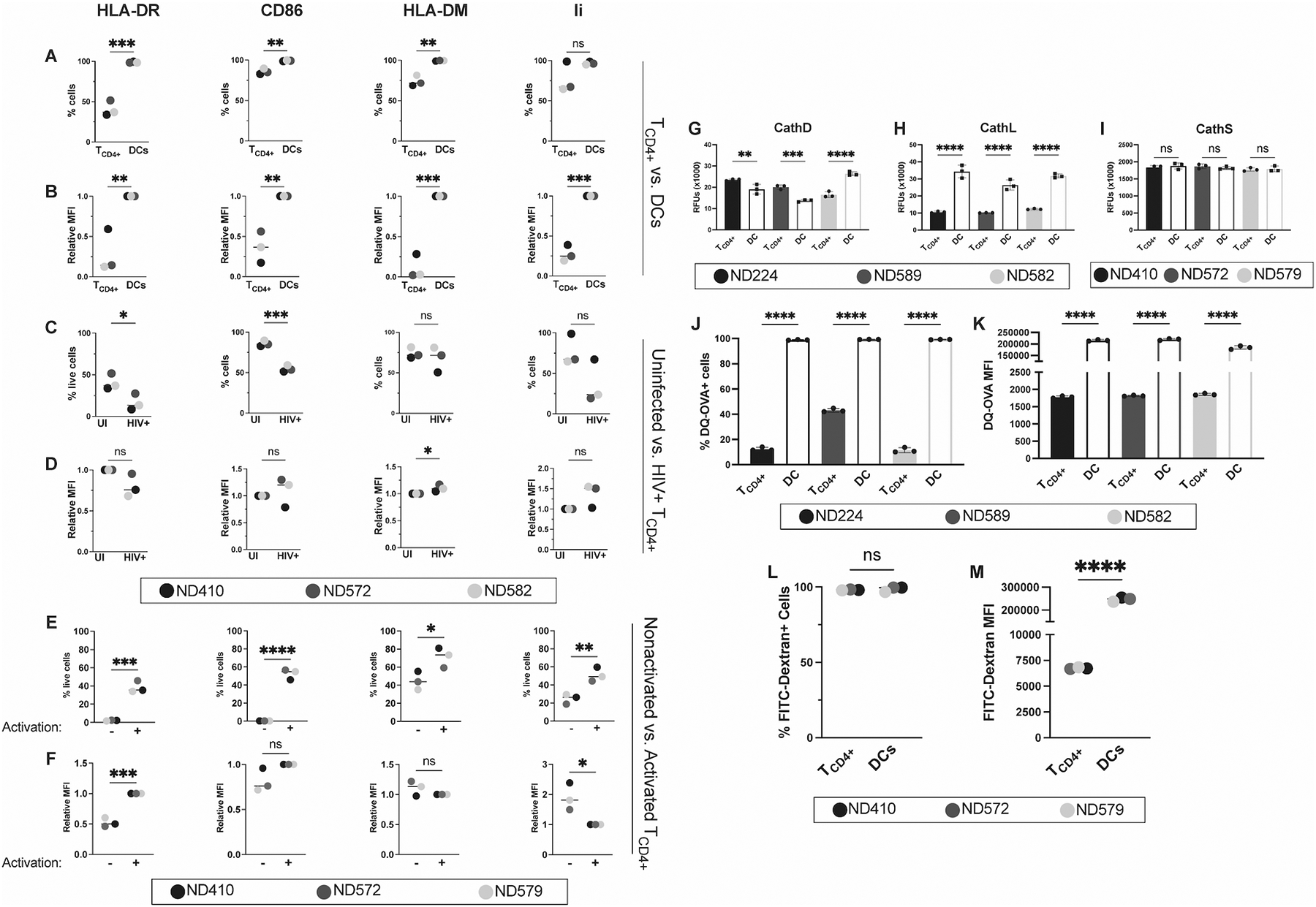
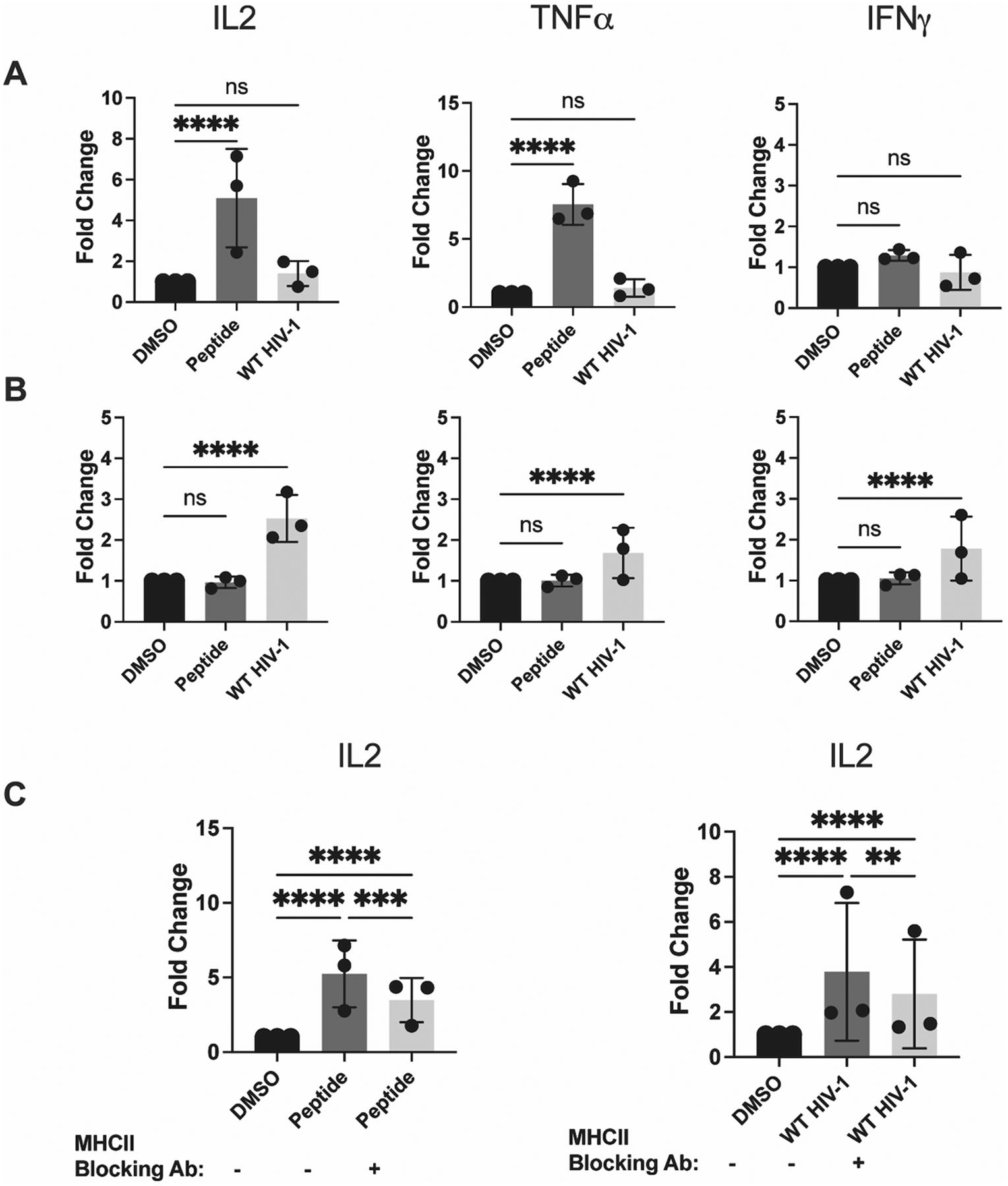
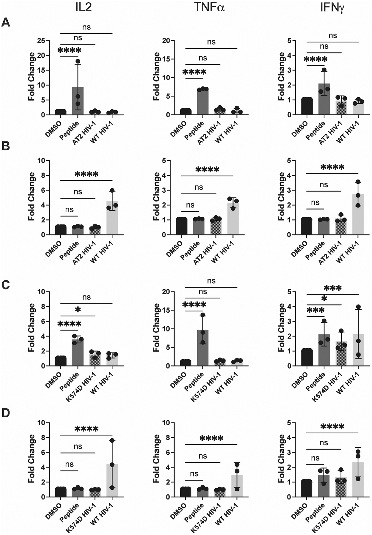
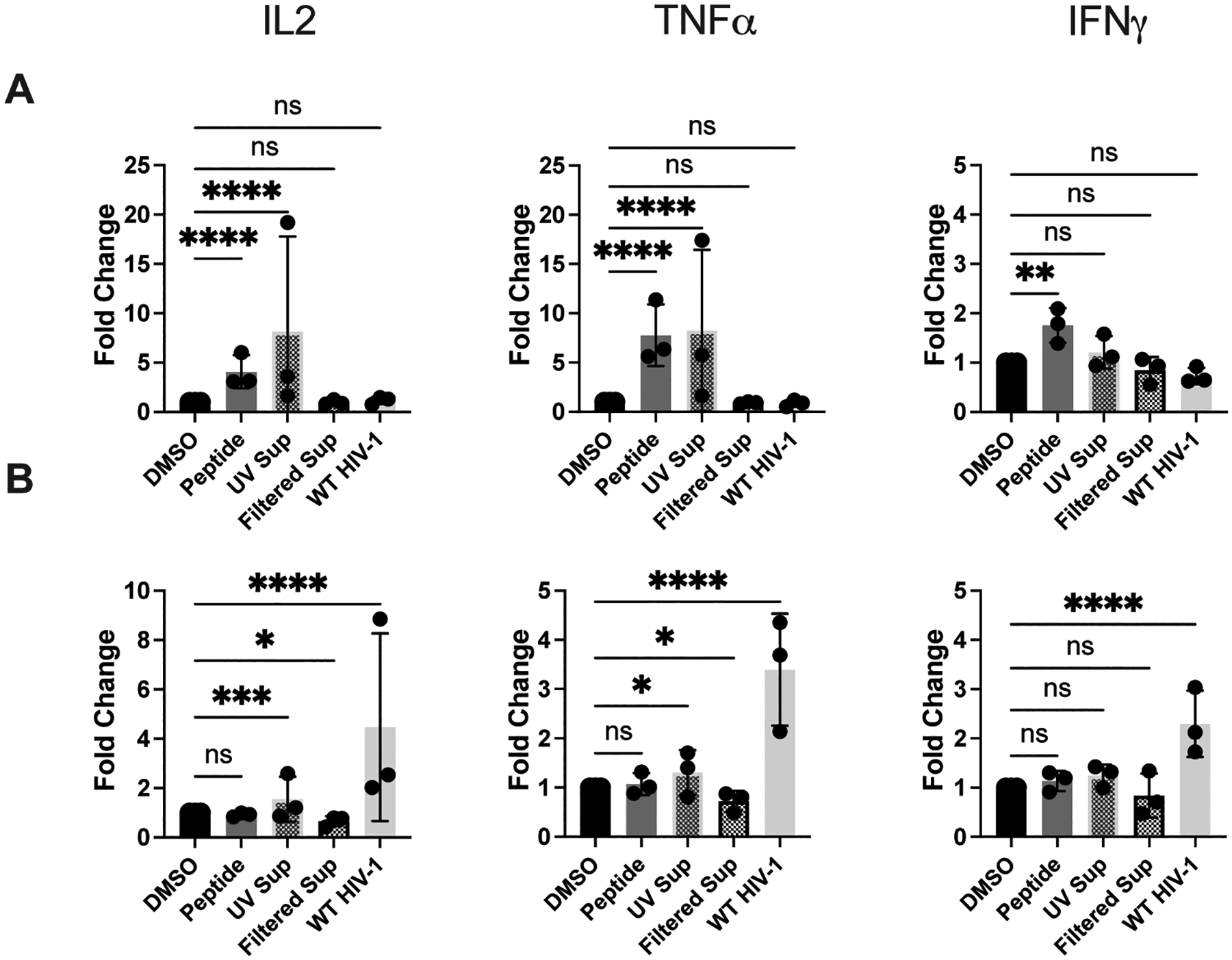
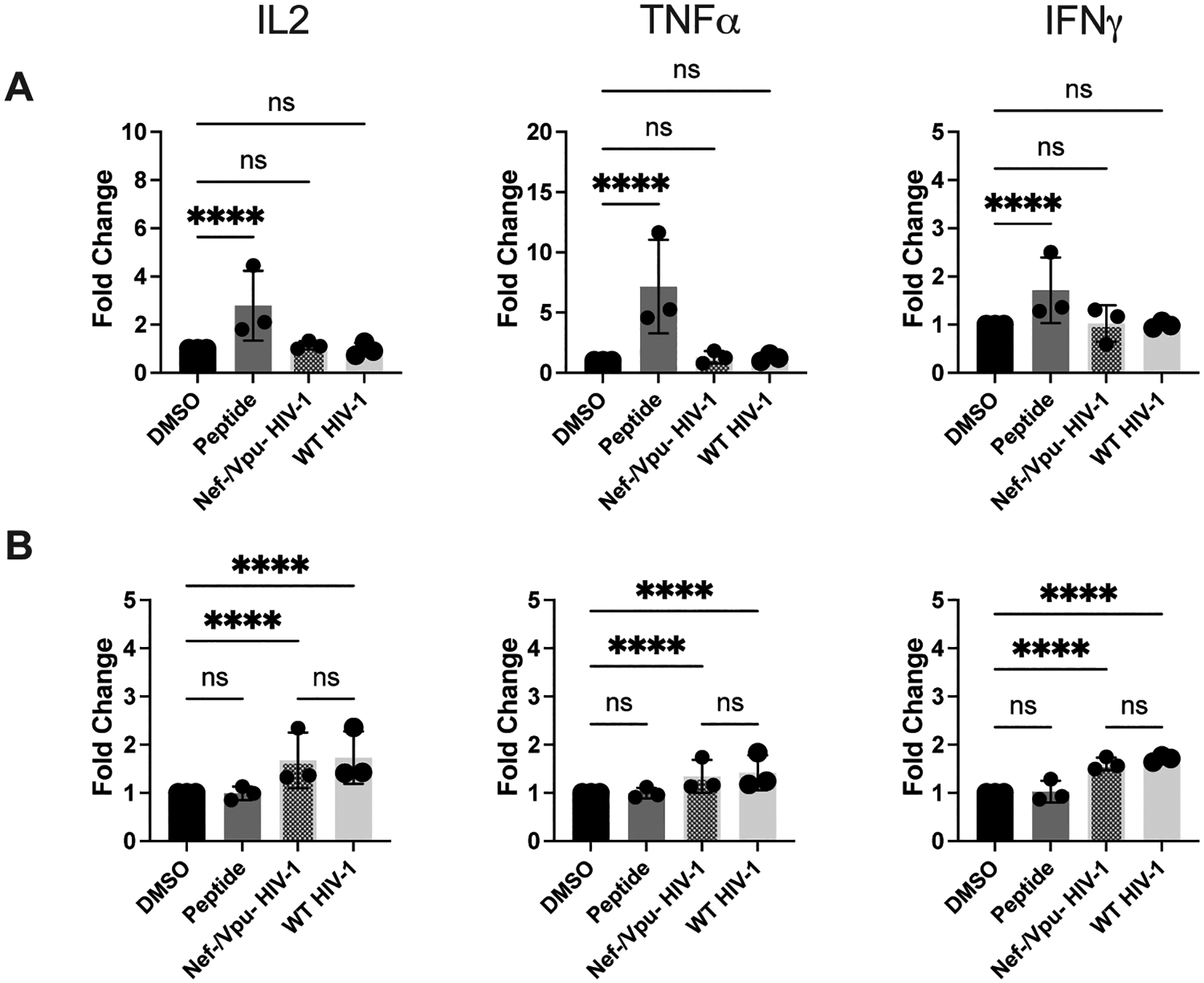
HIV-1-specific CD4+ T cells (TCD4+s) play a critical role in controlling HIV-1 infection. Canonically, TCD4+s are activated by peptides derived from extracellular ("exogenous") Ags displayed in complex with MHC class II (MHC II) molecules on the surfaces of "professional" APCs such as dendritic cells (DCs). In contrast, activated human TCD4+s, which express MHC II, are not typically considered for their APC potential because of their low endocytic capacity and the exogenous Ag systems historically used for assessment. Using primary TCD4+s and monocyte-derived DCs from healthy donors, we show that activated human TCD4+s are highly effective at MHC II-restricted presentation of an immunodominant HIV-1-derived epitope postinfection and subsequent noncanonical processing and presentation of endogenously produced Ag. Our results indicate that, in addition to marshalling HIV-1-specific immune responses during infection, TCD4+s also act as APCs, leading to the activation of HIV-1-specific TCD4+s.


Figures

Figure 1.
Figure 1.. Activated TCD4+ and DCs both express antigen presentation machinery components and have endocytic protease activities but TCD4+ have weak internalization capabilities.
(A) percent positive and (B) relative mean fluorescence intensity (MFI) of HLA-DR, CD86, HLA-DM, and invariant chain in uninfected TCD4+ and DCs. TCD4+ and DCs were cultured as described. Cells were then stained for flow cytometry. HLA-DR and CD86 positive populations were gated on live singlets. HLA-DM and invariant chain expression was determined by pre-gating HLA-DR and CD86 double positive cells. (C) Percent positive and (D) relative MFI of these markers in uninfected (UI) and WT HIV-1 infected (HIV+) TCD4+. Where indicated, activated TCD4+ were infected with 100 ng p24 HIV-1 3 days prior to staining. Activated TCD4+ and DCs lysates were then analyzed for (E) cathepsin D, (F) cathepsin L, and (G) cathepsin S activity using fluorometric cathepsin substrates. Lysates from 1 × 10⁵ cells were analyzed per replicate. Measurements were taken after 30 mins at 37°C. Activated TCD4+ and DCs were then tested for their ability to internalize and proteolyze DQ-OVA. All values are background subtracted. (H) Percent DQ-OVA positive cells and (I) MFI in DQ-OVA+ cells. TCD4+ and DCs were cultured as previously described and incubated with DQ-OVA fluorescent substrate for 2 hours at 37°C. Uptake and proteolysis were measured by flow cytometry on live singlets. Gates were drawn based on no-substrate controls. Percent FITC-Dextran positive cells (J) and (K) MFI in FITC-Dextran+ cells. TCD4+ and DCs were cultured as previously described and incubated with FITC-Dextran fluorescent substrate for 2 hours at 37°C. Uptake and proteolysis were measured by flow cytometry on live singlets. Gates were drawn based on no-substrate controls. Each color represents a unique donor, as indicated in the figure legend. For G-K, each dot represents a technical replicate. Representative of 3 independent experiments with the exception of L and M, which are representative of 2 independent experiments. For A-F and L-M, data were analyzed with an unpaired t test. For G-K, data were analyzed with a One-way ANOVA. Bars represent mean ± SD. *p<0.05, **p<0.01, ***p<0.005, ****p<0.001.
Figure 2.
Figure 2.. Activated TCD4+, but not DCs, can present epitope derived from infectious HIV-1 in an MHCII-dependent manner.
DCs and activated TCD4+ were cultured and infected as described in Materials and Methods. (A) DCs and (B) TCD4+ were assessed for their ability to present peptide and WT HIV-1 after 8 hours of coculture with HIV-1-specific TCD4+. IL-2, TNFα, and IFNγ expression was evaluated by flow cytometry. (C) DCs (left) and activated TCD4+ (right) were treated with 50 μg/mL MHCII blocking antibody 2 hours prior to the beginning of the assay. IL-2 expression was assessed by flow cytometry. Fold induction of each cytokine is shown, using DMSO as a baseline. Each dot represents an independent experiment. Bars represent mean ± SD. One way ANOVA, **p<0.01, ****p<0.001.
Figure 3.
Figure 3.. DCs and activated TCD4+ are unable to present epitope derived from AT2-inactivated and K574D fusion-deficient HIV-1.
DCs and activated TCD4+ were cultured and infected with WT HIV-1 as described in Materials and Methods. AT2-treated HIV-1 and K574D fusion-deficient HIV-1 were added to DCs and activated TCD4+ 12 hours prior to the beginning of the assay. (A) DCs and (B) TCD4+ were assessed for their ability to present AT2-treated HIV-1 after 8 hours of coculture with HIV-1-specific TCD4+. IL-2, TNFα, and IFNγ expression was assessed by flow cytometry. (C) DCs and (D) activated TCD4+ were assessed for their ability to present fusion-deficient HIV-1 in a similar manner. Fold induction of each cytokine is shown, using DMSO as a baseline. Each dot represents an independent experiment. Bars represent mean ± SD. One way ANOVA, *p<0.05, **p<0.01, ****p<0.001.
Figure 4.
Figure 4.. DCs, but not activated TCD4+, indirectly present HIV-1-derived epitope.
Presentation of UV-treated and filtered HIV-1+ supernatant by DCs and activated TCD4+ to HIV-1-specific TCD4+. DCs and activated TCD4+ were cultured and infected with WT HIV-1 as described in Materials and Methods. Supernatant from WT HIV-1-infected TCD4+ was collected and treated with UV light or filtered through a 100 kDa filter and then added to DCs and activated TCD4+ 12 hours prior to the beginning of the assay. (A) DCs and (B) TCD4+ were cocultured with HIV-1-specific TCD4+ for 8 hours and IL-2, TNFα, and IFNγ expression was assessed by flow cytometry. Fold induction of each cytokine is shown, using DMSO as a baseline. Each dot represents an independent experiment. Bars represent mean ± SD. One way ANOVA, *p<0.05, **p<0.01, ***p<0.005, ****p<0.001.
Figure 5.
Figure 5.. The viral accessory proteins Nef and Vpu do not impact the presentation of the Gag293 epitope by DCs and activated TCD4+.
DCs and activated TCD4+ were cultured and infected with WT HIV-1 and Nef-/Vpu- HIV-1 as described in Materials and Methods. (A) DCs and (B) TCD4+ were cocultured with HIV-1-specific TCD4+ for 8 hours and IL-2, TNFα, and IFNγ expression was assessed by flow cytometry. Fold induction of each cytokine is shown, using DMSO as a baseline. Each dot represents an independent experiment. Bars represent mean ± SD. One way ANOVA, ****p<0.001.

References

    1. UNAIDS. 2021. Global HIV & AIDS statistics — Fact sheet.
    1. Wilen CB, Tilton JC, and Doms RW. 2012. HIV: cell binding and entry. Cold Spring Harb Perspect Med 2. - PMC - PubMed
    1. Wallet C, De Rovere M, Van Assche J, Daouad F, De Wit S, Gautier V, Mallon PWG, Marcello A, Van Lint C, Rohr O, and Schwartz C. 2019. Microglial Cells: The Main HIV-1 Reservoir in the Brain. Front Cell Infect Microbiol 9: 362. - PMC - PubMed
    1. Raynaud-Messina B, Bracq L, Dupont M, Souriant S, Usmani SM, Proag A, Pingris K, Soldan V, Thibault C, Capilla F, Al Saati T, Gennero I, Jurdic P, Jolicoeur P, Davignon JL, Mempel TR, Benichou S, Maridonneau-Parini I, and Verollet C. 2018. Bone degradation machinery of osteoclasts: An HIV-1 target that contributes to bone loss. Proc Natl Acad Sci U S A 115: E2556–E2565. - PMC - PubMed
    1. Moir S, Chun TW, and Fauci AS. 2011. Pathogenic mechanisms of HIV disease. Annu Rev Pathol 6: 223–248. - PubMed

Publication types