. 2022 Oct:84:104270.
doi: 10.1016/j.ebiom.2022.104270. Epub 2022 Sep 18.

SARS-CoV-2 Omicron BA.5: Evolving tropism and evasion of potent humoral responses and resistance to clinical immunotherapeutics relative to viral variants of concern

Affiliations

SARS-CoV-2 Omicron BA.5: Evolving tropism and evasion of potent humoral responses and resistance to clinical immunotherapeutics relative to viral variants of concern

Anupriya Aggarwal et al. EBioMedicine. 2022 Oct.

Abstract

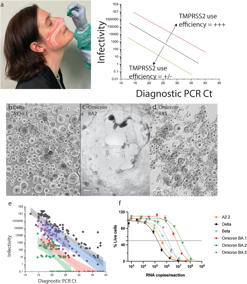
Background: Genetically distinct viral variants of severe acute respiratory syndrome coronavirus 2 (SARS-CoV-2) have been recorded since January 2020. The introduction of global vaccine programs has contributed to lower COVID-19 hospitalisation and mortality rates, particularly in developed countries. In late 2021, Omicron BA.1 emerged, with substantially altered genetic differences and clinical effects from other variants of concern. Shortly after dominating global spread in early 2022, BA.1 was supplanted by the genetically distinct Omicron lineage BA.2. A sub-lineage of BA.2, designated BA.5, presently has an outgrowth advantage over BA.2 and other BA.2 sub-lineages. Here we study the neutralisation of Omicron BA.1, BA.2 and BA.5 and pre-Omicron variants using a range of vaccine and convalescent sera and therapeutic monoclonal antibodies using a live virus neutralisation assay. Using primary nasopharyngeal swabs, we also tested the relative fitness of BA.5 compared to pre-Omicron and Omicron viral lineages in their ability to use the ACE2-TMPRSS2 pathway.

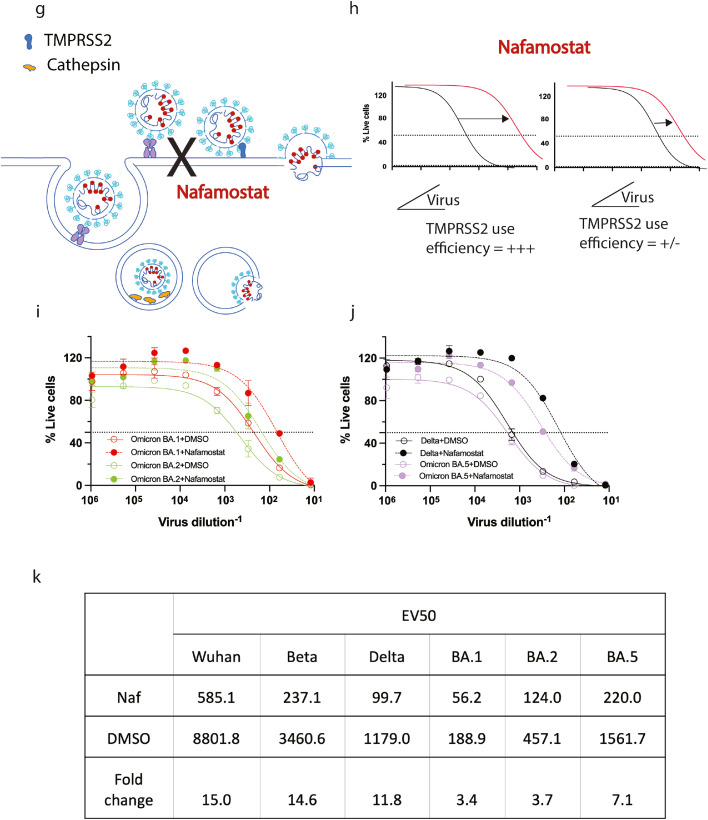
Methods: Using low passage clinical isolates of Clade A.2.2, Beta, Delta, BA.1, BA.2 and BA.5, we determined humoral neutralisation in vitro in vaccinated and convalescent cohorts, using concentrated human IgG pooled from thousands of plasma donors, and licensed monoclonal antibody therapies. We then determined infectivity to particle ratios in primary nasopharyngeal samples and expanded low passage isolates in a genetically engineered ACE2/TMPRSS2 cell line in the presence and absence of the TMPRSS2 inhibitor Nafamostat.

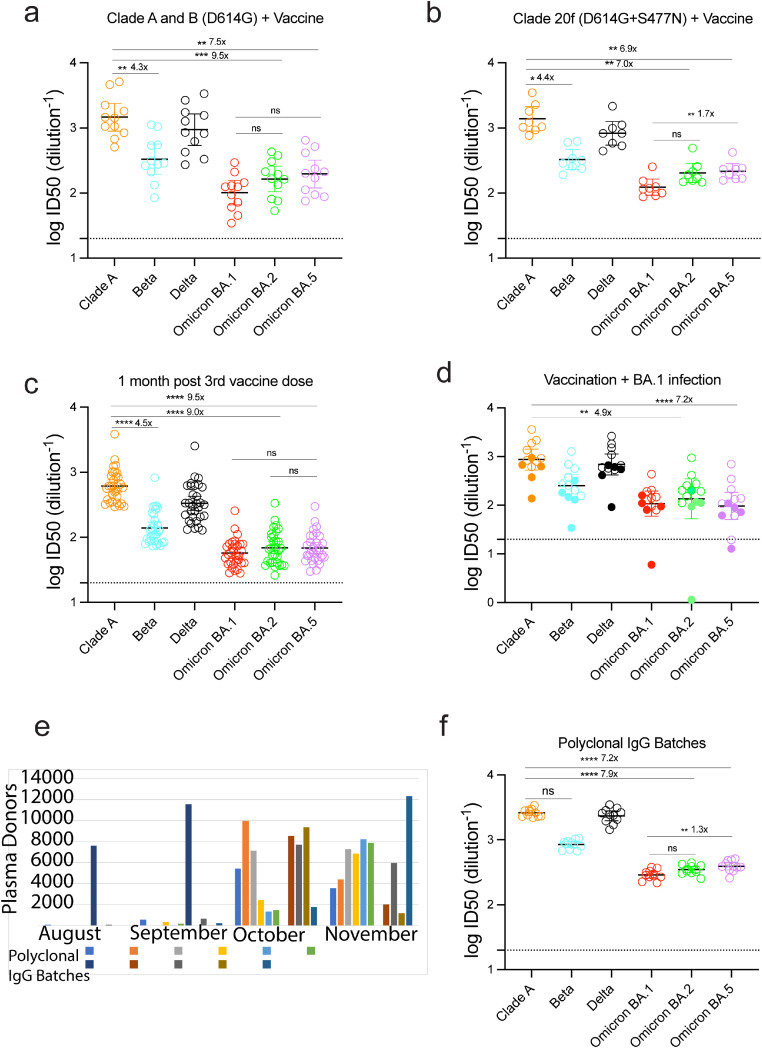
Findings: Peak responses to 3 doses of BNT162b2 vaccine were associated with a 9-fold reduction in neutralisation for Omicron lineages BA.1, BA.2 and BA.5. Concentrated pooled human IgG from convalescent and vaccinated donors and BNT162b2 vaccination with BA.1 breakthrough infections were associated with greater breadth of neutralisation, although the potency was still reduced 7-fold across all Omicron lineages. Testing of clinical grade antibodies revealed a 14.3-fold reduction using Evusheld and 16.8-fold reduction using Sotrovimab for the BA.5. Whilst the infectivity of BA.1 and BA.2 was attenuated in ACE2/TMPRSS2 entry, BA.5 was observed to be equivalent to that of an early 2020 circulating clade and had greater sensitivity to the TMPRSS2 inhibitor Nafamostat.

Interpretation: Observations support all Omicron variants to significantly escape neutralising antibodies across a range of vaccination and/or convalescent responses. Potency of therapeutic monoclonal antibodies is also reduced and differs across Omicron lineages. The key difference of BA.5 from other Omicron sub-variants is the reversion in tropism back to using the well-known ACE2-TMPRSS2 pathway, utilised efficiently by pre-Omicron lineages. Monitoring if these changes influence transmission and/or disease severity will be key for ongoing tracking and management of Omicron waves globally.

Funding: This work was primarily supported by Australian Medical Foundation research grants MRF2005760 (ST, GM & WDR), MRF2001684 (ADK and ST) and Medical Research Future Fund Antiviral Development Call grant (WDR), Medical Research Future Fund COVID-19 grant (MRFF2001684, ADK & SGT) and the New South Wales Health COVID-19 Research Grants Round 2 (SGT).

Keywords: ACE2; BA.2; BA.5; Neutralising antibodies; Omicron BA.1; SARS-CoV-2; TMPRSS2.


Conflict of interest statement

Declaration of interests A.O.S contributed to this manuscript in his capacity as adjunct associate lecturer (from January 2022) at the University of New South Wales. Since January 2022, A.O.S is an employee of GlaxoSmithKline Australia (medical science liaison for COVID-19).

Figures

Figure 1
Figure 1
Humoral neutralisation of clinical SARS-CoV-2 variants in convalescent and vaccinated donors, and pooled concentrated human IgG plasma samples. Neutralisation assays were performed in a high-throughput format in HAT-24 cells using live virus isolates from the variants of concern Delta (B.1.617.2), Beta (B.1.351), Omicron BA.1, Omicron BA.2 and Omicron BA.5 and the ancestral Wuhan-like virus with the original D614 background (A.2.2) as a control. ID50 neutralisation titres presented for 6 live variants for vaccinated donors from (a) Clade A and B (D614G) (First wave; n=10), (b) Clade 20F (D614G + S477N) (Second wave; n=7), (c) Healthy donors one month after third dose of vaccination (n=31), (d) Vaccinated (Open circles; n=9) or unvaccinated donors (Solid circles; n=5) infected with Omicron BA.1, (e) Number of United States based plasma donors per month sourced for polyclonal immunoglobulins (Poly-Ig; on average, greater than 10,000 donors were pooled for each batch tested) and (f) Concentrated polyclonal IgG from either convalescent and vaccinated donors. Data in (a-d and f) indicates the mean ID50 of technical replicates for individual samples (Circles) with the geometric mean and 95% confidence interval shown for each variant. Dotted lines show the limit of detection (LOD). Fold change reductions in ID50 neutralisation titres compare variants of concern to the ancestral variant and Omicron BA.1 where indicated. *p<0.05, **p<0.01, ***p<0.001, ****p<0.0001 for Kruskal Wallis test with Dunn's multiple comparison test. Data in (g-k) show the neutralisation activity of monoclonal antibodies (g) Sotrovimab, (h) Cilgavimab and Tixagevimab cocktail, (i) Cilgavimab alone and (j) Tixagevimab alone, against ancestral (k) A.2.2, Beta, Delta, Omicron BA.1, Omicron BA.2 and Omicron BA.5. IC50 values (ng/µL) and fold change are relative to ancestral A.2.2 for all monoclonal antibodies against Omicron BA.1, BA.2 and BA.5. Antibodies used herein were clinical grade batches.
Figure 1
Figure 1
Humoral neutralisation of clinical SARS-CoV-2 variants in convalescent and vaccinated donors, and pooled concentrated human IgG plasma samples. Neutralisation assays were performed in a high-throughput format in HAT-24 cells using live virus isolates from the variants of concern Delta (B.1.617.2), Beta (B.1.351), Omicron BA.1, Omicron BA.2 and Omicron BA.5 and the ancestral Wuhan-like virus with the original D614 background (A.2.2) as a control. ID50 neutralisation titres presented for 6 live variants for vaccinated donors from (a) Clade A and B (D614G) (First wave; n=10), (b) Clade 20F (D614G + S477N) (Second wave; n=7), (c) Healthy donors one month after third dose of vaccination (n=31), (d) Vaccinated (Open circles; n=9) or unvaccinated donors (Solid circles; n=5) infected with Omicron BA.1, (e) Number of United States based plasma donors per month sourced for polyclonal immunoglobulins (Poly-Ig; on average, greater than 10,000 donors were pooled for each batch tested) and (f) Concentrated polyclonal IgG from either convalescent and vaccinated donors. Data in (a-d and f) indicates the mean ID50 of technical replicates for individual samples (Circles) with the geometric mean and 95% confidence interval shown for each variant. Dotted lines show the limit of detection (LOD). Fold change reductions in ID50 neutralisation titres compare variants of concern to the ancestral variant and Omicron BA.1 where indicated. *p<0.05, **p<0.01, ***p<0.001, ****p<0.0001 for Kruskal Wallis test with Dunn's multiple comparison test. Data in (g-k) show the neutralisation activity of monoclonal antibodies (g) Sotrovimab, (h) Cilgavimab and Tixagevimab cocktail, (i) Cilgavimab alone and (j) Tixagevimab alone, against ancestral (k) A.2.2, Beta, Delta, Omicron BA.1, Omicron BA.2 and Omicron BA.5. IC50 values (ng/µL) and fold change are relative to ancestral A.2.2 for all monoclonal antibodies against Omicron BA.1, BA.2 and BA.5. Antibodies used herein were clinical grade batches.
Figure 2
Figure 2
Changing tropism of the BA.5 variant relative to BA.1, BA.2 and pre-Omicron variants. (a) Shows the shift in linear regression between virus infectivity and diagnostic PCR Ct values gives a measure of TMPRSS2 use by individual variants. Photo in (a) is not a participant in this study and consents to the inclusion of this photo. (b) Schematic showing the effect of TMPRSS2 inhibitor Nafamostat on virus entry at the cell membrane. (c) Efficiency of TMPRSS2 usage by the virus can be determined by observing a shift in VE50 when virus is titrated in the presence of saturating levels of Nafamostat. (d-f) Primary nasopharyngeal swabs were used to inoculate the HAT-24 cell line. All swabs are representative of high viral loads from diagnostic PCR Ct values of 17 to 19. Cultures of (d) AY39.1 (one of the last detected Delta lineages), (e) BA.2 and (f) BA.5 imaged 72 hours post-infection. Of note, only BA.2 and BA.5 were from samples collected at the same time period. Scale bars in d-f, 100 µm. Images in (d-f) are representative of >20 primary samples with high viral loads. Data in (f) is representative of three independent low passage expansions of the 6 isolates used. (g) Infectivity (TCID50/mL) is presented against original diagnostic PCR Ct values for BA.2 and BA.5. Shading represents the 95% confidence intervals for each linear regression. As a comparison, earlier tested samples from Delta and an early 2020 circulating clade are also presented. (h) Three early clade variants (A.2.2, Beta and Delta) and three Omicron sub-lineages were grown within a 24-hour time frame under identical culture conditions (i.e. expanded at the same time, with the same MOI and harvest time). Virion particle counts were then determined using quantitative RT-PCR. Titres were then determined overnight using the R-20 assay to establish infectivity per variant per viral particle. In brief, the HAT-24 cell line rapidly develops cytopathic effects overnight, in a dose-dependent manner that can be enumerated by nuclei counts using high-content microscopy. Infectivity can then be determined by calculating 50% death of cells within the culture (VE50). Each point and error bar represent the mean and standard deviation respectively of four technical replicates. Virus titrations of (i) BA.1, BA.2 and (j) Delta, BA.5 were carried out in the presence of 20 µM Nafamostat (Dashed line) or DMSO (Solid line) and (k) change in VE50 values was used as a measure of TMPRSS2 sensitivity. Data is representative of two independent experiments (N = 2).
Figure 2
Figure 2
Changing tropism of the BA.5 variant relative to BA.1, BA.2 and pre-Omicron variants. (a) Shows the shift in linear regression between virus infectivity and diagnostic PCR Ct values gives a measure of TMPRSS2 use by individual variants. Photo in (a) is not a participant in this study and consents to the inclusion of this photo. (b) Schematic showing the effect of TMPRSS2 inhibitor Nafamostat on virus entry at the cell membrane. (c) Efficiency of TMPRSS2 usage by the virus can be determined by observing a shift in VE50 when virus is titrated in the presence of saturating levels of Nafamostat. (d-f) Primary nasopharyngeal swabs were used to inoculate the HAT-24 cell line. All swabs are representative of high viral loads from diagnostic PCR Ct values of 17 to 19. Cultures of (d) AY39.1 (one of the last detected Delta lineages), (e) BA.2 and (f) BA.5 imaged 72 hours post-infection. Of note, only BA.2 and BA.5 were from samples collected at the same time period. Scale bars in d-f, 100 µm. Images in (d-f) are representative of >20 primary samples with high viral loads. Data in (f) is representative of three independent low passage expansions of the 6 isolates used. (g) Infectivity (TCID50/mL) is presented against original diagnostic PCR Ct values for BA.2 and BA.5. Shading represents the 95% confidence intervals for each linear regression. As a comparison, earlier tested samples from Delta and an early 2020 circulating clade are also presented. (h) Three early clade variants (A.2.2, Beta and Delta) and three Omicron sub-lineages were grown within a 24-hour time frame under identical culture conditions (i.e. expanded at the same time, with the same MOI and harvest time). Virion particle counts were then determined using quantitative RT-PCR. Titres were then determined overnight using the R-20 assay to establish infectivity per variant per viral particle. In brief, the HAT-24 cell line rapidly develops cytopathic effects overnight, in a dose-dependent manner that can be enumerated by nuclei counts using high-content microscopy. Infectivity can then be determined by calculating 50% death of cells within the culture (VE50). Each point and error bar represent the mean and standard deviation respectively of four technical replicates. Virus titrations of (i) BA.1, BA.2 and (j) Delta, BA.5 were carried out in the presence of 20 µM Nafamostat (Dashed line) or DMSO (Solid line) and (k) change in VE50 values was used as a measure of TMPRSS2 sensitivity. Data is representative of two independent experiments (N = 2).

References

    1. Rajah MM, Hubert M, Bishop E, et al. SARS-CoV-2 Alpha, Beta, and Delta variants display enhanced Spike-mediated syncytia formation. EMBO J. 2021;40(24) - PMC - PubMed
    1. Zhao H, Lu L, Peng Z, et al. SARS-CoV-2 Omicron variant shows less efficient replication and fusion activity when compared with Delta variant in TMPRSS2-expressed cells. Emerg Microbes Infect. 2022;11(1):277–283. - PMC - PubMed
    1. Meng B, Abdullahi A, Ferreira I, et al. Altered TMPRSS2 usage by SARS-CoV-2 Omicron impacts infectivity and fusogenicity. Nature. 2022;603(7902):706–714. - PMC - PubMed
    1. Halfmann PJ, Iida S, Iwatsuki-Horimoto K, et al. SARS-CoV-2 Omicron virus causes attenuated disease in mice and hamsters. Nature. 2022;603(7902):687–692. - PMC - PubMed
    1. Suzuki R, Yamasoba D, Kimura I, et al. Attenuated fusogenicity and pathogenicity of SARS-CoV-2 Omicron variant. Nature. 2022;603(7902):700–705. - PMC - PubMed

Supplementary concepts