Surgical management of Grisel syndrome in the adult patient: illustrative case
- PMID: 36130538
- PMCID: PMC9379629
- DOI: 10.3171/CASE21692
Surgical management of Grisel syndrome in the adult patient: illustrative case
Abstract
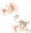
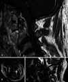
Background: Grisel syndrome describes an infectious soft tissue process that destabilizes the cervical bony elements and ligamentous complexes. This nontraumatic atlantoaxial rotary subluxation occurs in children primarily. This case illustrates a rare case presentation of an adult with Grisel syndrome: infectious destruction of the right atlantoaxial facet joint caused the occiput-C1 vertebra (head) to rotate rightward with lateral horizontal displacement off the C2 vertebra.
Observations: Because the infection destroyed the C1 bony arch and atlantoaxial facet joints with epidural extension, the rotated head and atlas pulled the brainstem-cervical spinal cord junction against a fixed odontoid process, resulting in a cord contusion. Because of the highly unstable craniocervical junction, the patient presented with torticollis and left upper extremity weakness.
Lessons: Treatment entailed closed reduction under general anesthesia followed by occipitocervical fusion with an occipital plate, C1 lateral mass screws, and C2-C5 pedicle screws. This case describes the unique surgical pearls necessary for occipitocervical fusion of an unstable craniocervical junction, including tips with neuronavigation, trajectories of the cervical pedicle screws, aligning the lateral mass and pedicle screws with the occipital plate, and nuances with occipitocervical distraction.
Keywords: Grisel syndrome; atlantoaxial; cervical; pedicle; rotary; subluxation; surgery.
Conflict of interest statement
Figures


Similar articles
-
Pediatric cervical kyphosis in the MRI era (1984-2008) with long-term follow up: literature review.Childs Nerv Syst. 2022 Feb;38(2):361-377. doi: 10.1007/s00381-021-05409-z. Epub 2021 Nov 22. Childs Nerv Syst. 2022. PMID: 34806157 Review.
-
Posterior occipitocervical reconstruction using cervical pedicle screws and plate-rod systems.Spine (Phila Pa 1976). 1999 Jul 15;24(14):1425-34. doi: 10.1097/00007632-199907150-00007. Spine (Phila Pa 1976). 1999. PMID: 10423787
-
Bilateral C1 laminar hooks combined with C2 pedicle screw fixation in the treatment of atlantoaxial subluxation after Grisel syndrome.Spine J. 2016 Dec;16(12):e755-e760. doi: 10.1016/j.spinee.2016.08.016. Epub 2016 Sep 23. Spine J. 2016. PMID: 27669669
-
Posterior screw placement on the lateral mass of atlas: an anatomic study.Spine (Phila Pa 1976). 2004 Mar 1;29(5):500-3. doi: 10.1097/01.brs.0000113874.82587.33. Spine (Phila Pa 1976). 2004. PMID: 15129062
-
Fusions at the craniovertebral junction.Childs Nerv Syst. 2008 Oct;24(10):1209-24. doi: 10.1007/s00381-008-0607-7. Epub 2008 Apr 4. Childs Nerv Syst. 2008. PMID: 18389260 Review.
Cited by
-
Torticollis in a child with Grisel syndrome: A case report and review of the literature.Int J Surg Case Rep. 2025 Feb;127:110817. doi: 10.1016/j.ijscr.2025.110817. Epub 2025 Jan 4. Int J Surg Case Rep. 2025. PMID: 39823970 Free PMC article.
References
-
- Grisel P. Felix Dévé. Presse Med. 1951;59(78):1647–1648. - PubMed
-
- Bissonnette B. Grisel syndrome. In: Bissonnette B, Luginbuehl I, Marciniak B, Dalens BJ, editors. Syndromes: Rapid Recognition and Perioperative Implications. McGraw-Hill; 2006. Accessed February 16, 2022. https://accessanesthesiology.mhmedical.com/content.aspx?bookid=852§i....
-
- Youssef K, Daniel S. Grisel syndrome in adult patients. Report of two cases and review of the literature. Can J Neurol Sci. 2009;36(1):109–113. - PubMed
-
- Agha RA, Sohrabi C, Mathew G, Franchi T, Kerwan A, O’Neill N. The PROCESS 2020 guideline: updating consensus Preferred Reporting of CasESeries in Surgery (PROCESS) guidelines. Int J Surg. 2020;84:231–235. - PubMed
LinkOut - more resources
Full Text Sources
Miscellaneous

