Clinical Trial
. 2022 Sep 21;13(1):5308.
doi: 10.1038/s41467-022-32944-3.

Preclinical and randomized clinical evaluation of the p38α kinase inhibitor neflamapimod for basal forebrain cholinergic degeneration

Affiliations
Clinical Trial

Preclinical and randomized clinical evaluation of the p38α kinase inhibitor neflamapimod for basal forebrain cholinergic degeneration

Ying Jiang et al. Nat Commun. .

Abstract

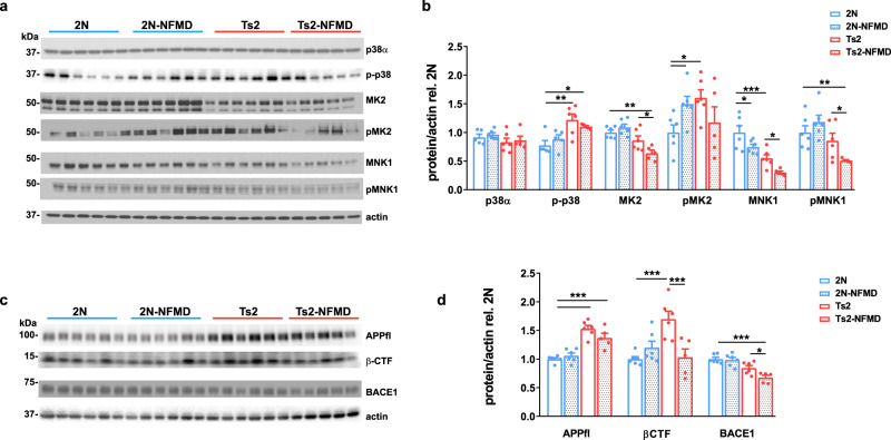
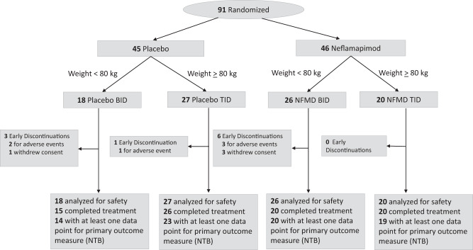
The endosome-associated GTPase Rab5 is a central player in the molecular mechanisms leading to degeneration of basal forebrain cholinergic neurons (BFCN), a long-standing target for drug development. As p38α is a Rab5 activator, we hypothesized that inhibition of this kinase holds potential as an approach to treat diseases associated with BFCN loss. Herein, we report that neflamapimod (oral small molecule p38α inhibitor) reduces Rab5 activity, reverses endosomal pathology, and restores the numbers and morphology of BFCNs in a mouse model that develops BFCN degeneration. We also report on the results of an exploratory (hypothesis-generating) phase 2a randomized double-blind 16-week placebo-controlled clinical trial (Clinical trial registration: NCT04001517/EudraCT #2019-001566-15) of neflamapimod in mild-to-moderate dementia with Lewy bodies (DLB), a disease in which BFCN degeneration is an important driver of disease expression. A total of 91 participants, all receiving background cholinesterase inhibitor therapy, were randomized 1:1 between neflamapimod 40 mg or matching placebo capsules (taken orally twice-daily if weight <80 kg or thrice-daily if weight >80 kg). Neflamapimod does not show an effect in the clinical study on the primary endpoint, a cognitive-test battery. On two secondary endpoints, a measure of functional mobility and a dementia rating-scale, improvements were seen that are consistent with an effect on BFCN function. Neflamapimod treatment is well-tolerated with no study drug associated treatment discontinuations. The combined preclinical and clinical observations inform on the validity of the Rab5-based pathogenic model of cholinergic degeneration and provide a foundation for confirmatory (hypothesis-testing) clinical evaluation of neflamapimod in DLB.


Conflict of interest statement

J.J.A, A.G., and K.B. are employees of EIP Pharma, the sponsor of the clinical study. J.J.A. is also founder of and has stock ownership in EIP Pharma. U.A.G. receives compensation as a scientific consultant to EIP Pharma. S.N.G. has served on Advisory Boards of Jannsen, Acadia, and Sanofi, has received consulting fees from EIP Pharma, and has received funding from the NIH, the DOD CDMRP, the Michael J. Fox Foundation, the FFFPRI, and the Lewy Body Dementia Association. N.D.P. is CEO and co-owner of Brain Research Center. P.M. is a full-time employee at Cogstate Ltd. J.E.H. reports receipt of personal fees in the past 2 years from Actinogen, AlzeCure, Aptinyx, Astra Zeneca, Athira Therapeutics, Axon Neuroscience, Axovant, Bial Biotech, Biogen Idec, BlackThornRx, Boehringer Ingelheim, Brands2life, Cerecin, Cognito, Cognition Therapeutics, Compass Pathways, Corlieve, Curasen, EIP Pharma, Eisai, G4X Discovery, GfHEU, Heptares, Ki Elements, Lundbeck, Lysosome Therapeutics, MyCognition, Neurocentria, Neurocog, Neurodyn Inc, Neurotrack, the NHS, Novartis, Novo Nordisk, Nutricia, Probiodrug, Prothena, Recognify, Regeneron, reMYND, Rodin Therapeutics, Samumed, Sanofi, Signant, Syndesi Therapeutics, Takeda, Vivoryon Therapeutics and Winterlight Labs. In addition, he holds stock options in Neurotrack Inc. and is a joint holder of patents with My Cognition Ltd. C.E.T. has a collaboration contracts with ADx Neurosciences, Quanterix and Eli Lilly; performed contract research or received grants from AC-Immune, Axon Neurosciences, Bioconnect, Bioorchestra, Brainstorm Therapeutics, Celgene, EIP Pharma, Eisai, Grifols, Novo Nordisk, PeopleBio, Roche, Toyama, and Vivoryon; and has had speaker contracts for Roche, Grifols, and Novo Nordisk. P.S. has received consultancy fees (paid to the institution) from AC Immune, Brainstorm Cell, EIP Pharma, ImmunoBrain Checkpoint, Genentech, Novartis, Novo Nordisk. P.S. is also principal investigator of studies with AC Immune, FUJI-film/Toyama, UCB, and Vivoryon; and is an employee of EQT Life Sciences (formerly LSP). The remaining authors declare no competing interests.

Figures

Fig. 1
Fig. 1. Normalized Rab5+ endosome number/size and restored cholinergic neuronal numbers in Ts2 mice treated with neflamapimod (NFMD).
a Representative images of medial septal nucleus (MSN) regions from vehicle-treated wildtype (2 N, n = 4), vehicle-treated Ts2 (n = 4) and NFMD-treated Ts2 (Ts2-NFMD) mice, labeled with Rab5-GTP (red) and Rab5a (green) antibodies; arrows point to the Rab5-positive neurons, which are shown enlarged in the lower panel (scale bar, 15 μm). b The numbers, sizes and areas of Rab5-GTP-positive endosomes determined by Image J analysis [n = 4 mice per group; for number: F(3,12) = 7.789, R square = 0.661; for 2 N vs Ts2 p = 0.0202, 95% CI = −1.880, −0.151; for Ts2 vs Ts2-NFMD p = 0.0097, 95% CI = 0.273, 2.002; for size: F(3,12) = 6.939, R square = 0.634, for 2 N vs Ts2 p = 0.0036, 95% CI = −1.329, −0.269; for area: F(3,12) = 13.19, R square = 0.767; for 2 N vs Ts2 p = 0.0009; 95% CI = −3.766, −1.079; for Ts2 vs Ts2-NFMD p = 0.0030, 95% CI = 0.7325, 3.419]. c The ratio of Rab5-GTP to total Rab5 intensities determined with Image J [n = 4 mice per group; F(3,12) = 13.23, R square = 0.768; for 2 N vs Ts2 p = 0.001, 95% CI = −0.537, −0.073; for Ts2 vs Ts2-NFMD p = 0.0002, 95% CI = 0.254, 0.719]. d The number of stereologically counted ChAT+ neurons in the MSN region of 2 N (n = 27) and Ts2 (n = 28) mice was graphed versus age of mice in months (Linear regression F = 10.16, R square = 0.281, p = 0.0037, 95% CI = −0.259, −0.056 for Ts2; F = 0.548, R square = 0.0215, p = 0.466, 95% CI = -infinity, −3.98) for 2N. e Representative images of diaminobenzidine (DAB)-stained ChAT+ neurons in the MSN region of 2 N and Ts2 mice treated with either vehicle or NFMD (scale bar, 100 μm). f Quantification of DAB-stained ChAT+ neurons in the MSN region; n = 10 (2 N), n = 11 (2N-NFMD), n = 14 (Ts2) and n = 11 (Ts2-NFMD) mice (F(3, 43) = 11.10, R square = 0.436, for 2 N vs Ts2, p < 0.0001, 95% CI = 0.588, 1.712; for Ts2 vs Ts2-NFMD p = 0.0037, 95% CI = −1.331, −207). g, h Abnormal morphology of ChAT+ neurons was consistently and reproducibly seen in the MSN of Ts2 mice, with swelling (yellow arrows), perikaryal atrophy (red arrows) and below normal ChAT immunoreactivity intensity (green arrows) in the representative images (scale bar,10 μm). Data are presented as mean values ± SEM. Graph made and analyzed with GraphPad Prism8.0.1 with Ordinary One-Way ANOVA/Tukey correction. Statistical significance is represented by asterisks *p ≤ 0.05, **p ≤ 0.01, ***p ≤ 0.001. Source data are provided as a Source Data File.
Fig. 2
Fig. 2. Slower decline of LTP and normalized scores of behavioral tests in Ts2 mice treated with NFMD.
a Input/output relationship plots of hippocampal slices from 2 N and Ts2 mice treated with either vehicle or NFMD (n = 4 mice for Ts2 group and n = 5 mice for the other treatment groups), and graphed using GraphPad Prism8.0.1 [no significant difference between the slopes, Linear regression F (3, 40) = 0.107, p = 0.956]. b Plots of LTP in the Schaffer collateral synapses (CA3-CA1) induced by theta-burst stimulation (TBS) of mice from four treatment groups [Linear regression for, significant difference between the slopes of 2 N vs Ts2, F(1, 236) = 6.925, p = 0.0091; no significant difference between the slopes of Ts2 vs Ts2-NFMD, F(1, 236) = 2.103, p = 0.148]. c Averages of fEPSP slopes at 1, 50, and 110 min following tetanic stimulation; NFMD treatment increased the fEPSP slope of Ts2 mice by 18% at the 110 min time point [Ordinary One-Way ANOVA with Tukey correction, at 1 min time point, F(3, 34) = 20.75, R square = 0.647, for 2 N vs Ts2, p < 0.0001, 95% CI = 54.62, 139.5; at 50 min time point, F(3, 34) = 16.44, R square = 0.592, for 2 N vs Ts2, p < 0.0001, 95% CI = 49.54, 144.5; at 110 min time point, F(3, 34) = 19.00, R square = 0.626, for 2 N vs Ts2, p < 0.0001, 95% CI = 72.09, 139.6, for Ts2 vs Ts2-NFMD, p = 0.0334, 95% CI = −67.71, −2.938]. d Novel Object Recognition (NOR) test at 24 h after familiarization session, represented by recognition index in 2 N (n = 8) and Ts2 (n = 8) mice before (Pre) and after (Post) 4 weeks of NFMD treatments [Ordinary One-Way ANOVA with Tukey correction, F(3, 24) = 1.967, R square = 0.197, for Ts2-pre vs Ts2-post, p = 0.0262, 95% CI = 2.406, 34.97]. e Open field test results including speed, distance and percentage of time spent in thigmotaxis for 2 N (n = 8) and Ts2 mice (n = 8) Pre and Post 4 weeks of NFMD treatment [Ordinary One-Way ANOVA with Tukey correction, For Speed F(3, 22) = 5.936, R square = 0.447, for 2N-NFMD-pre vs Ts2-NFMD-pre, p = 0.0148, 95% CI = −36.97, −4.47; for Ts2-NFMD-pre vs Ts2-NFMD-post p = 0.0148, 95% CI = 3.572, 34.89; For Distance F(3, 22) = 9.784, R square = 0.572, for 2N-NFMD-pre vs Ts2-NFMD-pre, p = 0.0434, 95% CI = −27.75, −0.295; for Ts2-NFMD-pre vs Ts2-NFMD-post p = 0.0003, 95% CI = 9.205, 32.77). Data are presented as mean values ± SEM. Statistical significance represented by asterisks *p ≤ 0.05, **p ≤ 0.01, ***p ≤ 0.001. Source data are provided as a Source Data File.
Fig. 3
Fig. 3. The positive effects of neflamapimod on pathology and function in Ts2 mice appear to be mediated by inhibition of the p38α kinase pathway and related to BACE1 and βCTF reductions.
a Western blot analysis of p38α, phosphorylated-p38 (p-p38), and downstream substrates MK2 and MNK1 in tissue homogenates of the brain cortex after 2 weeks of either vehicle or NFMD treatment (n = 5 for Ts2-NFMD, and n = 6 for other treatment groups). b Quantification of the western blot images shown with Image J and graphed with GraphPad Prism 8.0.1 [Ordinary One-Way ANOVA, For p-p38 F(3, 19) = 6.658, R square = 0.540, for 2 N vs Ts2, p = 0.0044, 95% CI = −0.760, −0.131; for 2 N vs Ts2-NFMD p = 0.0495, 95% CI = −0.656 −0.000194; For MK2 F(3, 19) = 10.33, R square = 0.610, for 2 N vs Ts2-NFMD, p = 0.0005, 95% CI = 0.182, 0.541; for Ts2 vs Ts2-NFMD p = 0.0166, 95% CI = 0.0457, 0.404; For pMK2 F(3, 19) = 2.850, R square = 0.310, for 2 N vs Ts2, p = 0.0178, 95% CI = −1.095, −0.117; for 2 N vs 2N-NFMD p = 0.0469, 95% CI = −0.985, −0.0076; for Ts2 vs Ts2-NFMD p = 0.0166, 95% CI = 0.0457, 0.404; For MNK1 F(3, 19) = 15.11, R square = 0.705, for 2 N vs Ts2, p = 0.0003, 95% CI = 0.235, 0.668; for 2 N vs 2N-NFMD p = 0.023, 95% CI = 0.0393, 0.472, for Ts2 vs Ts2-NFMD, p = 0.0347, 95% CI = 0.0197, 0.473; For pMNK1 F(3, 19) = 6.285, R square = 0.498, for 2 N vs Ts2-k NFMD, p = 0.0063, 95% CI = 0.156, 0.824; for TS2 vs Ts2-NFMD p = 0.0428, 95% CI = 0.125, 0.681]. c, d Subsequent western blot analysis for full length APP (APPfl), APP-βCTF and BACE1 and western blot image quantified with Image J and graphed with GraphPad Prism 8.0.1 (Ordinary One-Way ANOVA, For APPfl F(3, 19) = 23.33, R square = 0.786, for 2 N vs Ts2, p < 0.0001, 95% CI = −684, −0.376; for 2 N vs Ts2-NFMD p = 0.0001, 95% CI = −0.532, −0.209; For APP-βCTF F(3, 19) = 8.167, R square = 0.563, for 2 N vs Ts2, p = 0.0003, 95% CI = −1.027, −0.367; for Ts2 vs Ts2-NFMD p = 0.0007, 95% CI = 0.3187, 1.010; For BACE1 F(3, 19) = 12.86, R square = 0.670, for 2 N vs Ts2-NFMD, p = 0.0001, 95% CI = 0.158, 0.491; for Ts2 vs TS2-NFMD p = 0.0469, 95% CI = −0.00185, 0.335). Data are presented as mean values ± SEM. Statistical significance is represented by asterisks *p ≤ 0.05, **p ≤ 0.01, ***p ≤ 0.001. Source data are provided as a Source Data File.
Fig. 4
Fig. 4. CONSORT flow diagram for the AscenD-LB Phase 2a clinical trial.
CONSORT 2010 Statement flow diagram showing participant flow through each stage of the AscenD-LB phase 2a randomized controlled trial from randomization through to follow-up and data analysis.

References

    1. Grothe MJ, et al. Atrophy of the cholinergic basal forebrain in dementia with Lewy bodies and Alzheimer’s disease dementia. J. Neurol. 2014;261:1939–1948. doi: 10.1007/s00415-014-7439-z. - DOI - PubMed
    1. Duda JE. Pathology and neurotransmitter abnormalities of dementia with Lewy bodies. Dement Geriatr. Cogn. Disord. 2004;17:3–14. doi: 10.1159/000074677. - DOI - PubMed
    1. Schumacher, J. et al. Cholinergic white matter pathways in dementia with Lewy bodies and Alzheimer’s disease. Brain (2021). - PMC - PubMed
    1. Schumacher J, et al. In vivo nucleus basalis of Meynert degeneration in mild cognitive impairment with Lewy bodies. Neuroimage Clin. 2021;30:102604. doi: 10.1016/j.nicl.2021.102604. - DOI - PMC - PubMed
    1. Schmitz TW, Spreng RN, The-Alzheimer’s-Disease-Neuroimaging-Initiative. Basal forebrain degeneration precedes and predicts the cortical spread of Alzheimer’s pathology. Nat. Commun. 2016;7:13249. doi: 10.1038/ncomms13249. - DOI - PMC - PubMed

Publication types

MeSH terms

Substances

Associated data