. 2022 Sep 21;13(1):5544.
doi: 10.1038/s41467-022-33191-2.

A de novo matrix for macroscopic living materials from bacteria

Affiliations

A de novo matrix for macroscopic living materials from bacteria

Sara Molinari et al. Nat Commun. .

Abstract

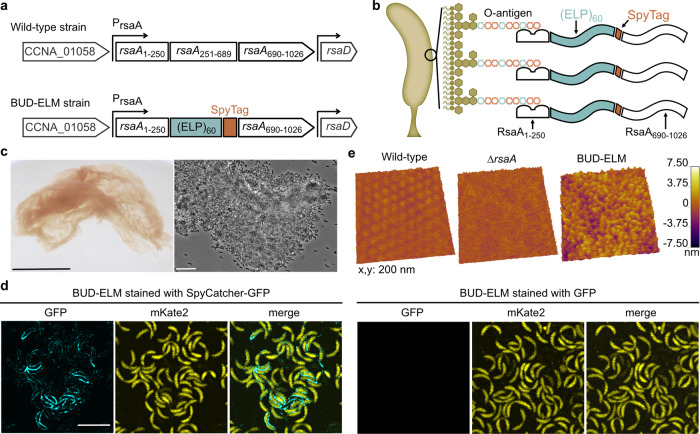
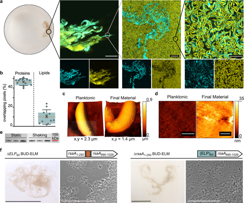
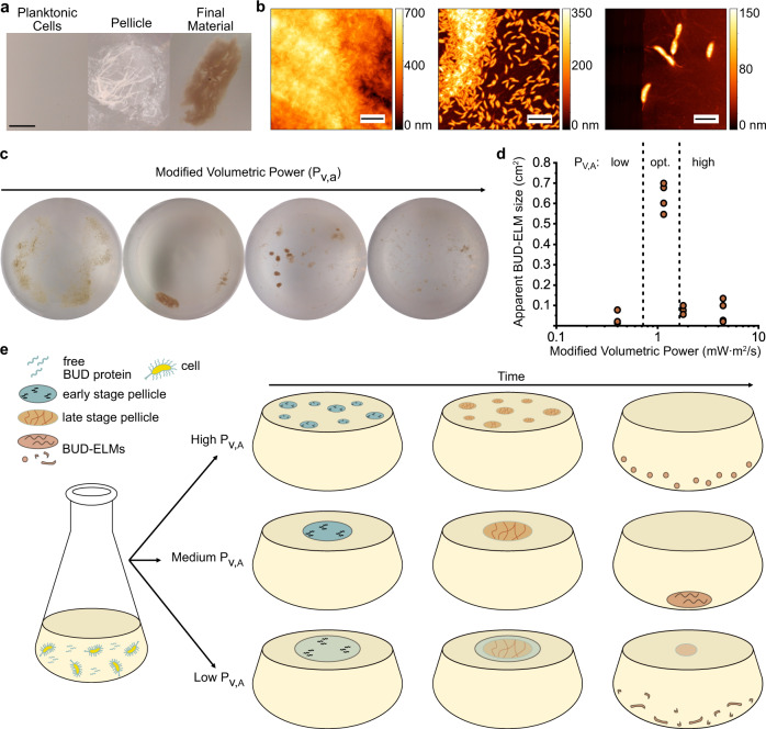
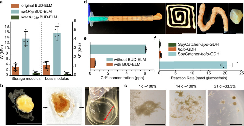
Engineered living materials (ELMs) embed living cells in a biopolymer matrix to create materials with tailored functions. While bottom-up assembly of macroscopic ELMs with a de novo matrix would offer the greatest control over material properties, we lack the ability to genetically encode a protein matrix that leads to collective self-organization. Here we report growth of ELMs from Caulobacter crescentus cells that display and secrete a self-interacting protein. This protein formed a de novo matrix and assembled cells into centimeter-scale ELMs. Discovery of design and assembly principles allowed us to tune the composition, mechanical properties, and catalytic function of these ELMs. This work provides genetic tools, design and assembly rules, and a platform for growing ELMs with control over both matrix and cellular structure and function.


Conflict of interest statement

The authors declare no competing interests.

Figures

Fig. 1
Fig. 1. Engineered strains of C. crescentus self-assemble into BUD-ELMs.
a Schematic of the native rsaA gene within its genomic context, showing its N-terminal cell anchoring domain (rsaA1–250) and C-terminal domain (rsaA251–1026) with the secretion subdomain (rsaA690–1026) (top). Schematic of the construct replacing the native rsaA gene in the BUD-ELM strain (bottom). b Illustration of the redesigned external surface of C. crescentus. showing the BUD protein attached to the cell surface. Absolute dimensions and relative positions of objects are only meant for illustrative purposes. c Photograph of free-floating material formed by the BUD-ELM strain (left); the scale bar is 1 cm. Brightfield image of a portion of a BUD-ELM (right), showing cell clusters and intact cells; scale bar is 10 µm. d Confocal microscopy of single cells of BUD-ELM strain stained with SpyCatcher-GFP (left) or GFP (right), demonstrating that the BUD protein is located on the cell surface. The scale bar is 5 µm and applies to every image. e AFM images of the cell surface of wild-type (left), ∆rsaA (middle), and BUD-ELM strain (right), showing the brush-like structure of the BUD-ELM strain’s surface.
Fig. 2
Fig. 2. BUD-ELMs contain a de novo protein matrix and display a hierarchical structure.
a Confocal microscopy of ELMs stained with SpyCatcher-GFP at increasing magnifications, showing a hierarchical structure. The bottom images show individual fluorescent channels: GFP (matrix) on the left and mKate2 (cells) on the right. Scale bars are, from left to right, 100, 10, and 5 µm, respectively. b Percentage of overlapping pixels between cell-free and stained regions, confirming the absence of lipids in the BUD protein matrix. Error bars are centered on the mean value (cross) and represent the standard deviation; the red line indicates the median value; the boxes show the interquartile range (25–75%). The analysis has been performed on 16 images for the protein and 11 for the lipid staining, each obtained from three independent samples. Source data are provided as a Source Data file. c AFM images of single cells at early (left) and late (right) stages of BUD-ELM formation, showing a difference in surface morphology. d High-resolution AFM images of single-cell surfaces at early (left) and late (right) stages of BUD-ELM formation, showing differences in surface layer thickness. Scale bars are 100 nm. e Immunoblot of BUD protein was detected in the growth media of culture grown in static (left) and shaking (right) conditions, showing a similar amount of protein in both conditions. BUD proteins were stained with the ANTI-FLAG® antibody. Source data are provided as a Source Data file. f Comparison between the ∆ELP60 and ∆rsaA1–250 BUD-ELM strains, showing differences in morphology and cell content. Each panel shows the genetic constructs (top), a representative image of BUD-ELMs at low (bottom, left), and high (bottom, right) magnification. For each panel, scale bars are 1 cm (bottom, left) and 50 µm (bottom, right).
Fig. 3
Fig. 3. BUD-ELMs are formed through a shaking-dependent, multi-step process.
a Optical images of representative BUD-ELM strain culture during material formation, showing BUD-ELMs are formed through a multi-step process. The scale bar is 1 cm. b AFM images of pellicle structure, showing the pellicle contains both a central region containing several layers of densely packed cells (left) and a peripheral region containing sparse cells connected by a thin membrane (center and right). Scale bars are, from left to right, 5, 5, and 2 µm, respectively. c Representative optical images of BUD-ELMs grown in 250 mL flasks under different modified volumetric power values. Altering the modified volumetric power changes the morphology and size of BUD-ELMs. d Correlation between modified volumetric power and the apparent surface area of BUD-ELMs grown in 500 mL shake flasks. Dotted lines separate the three ranges of PV,A: low, optimal (intermediate) and high. The graph shows that the apparent surface of BUD-ELMs grown in a 500 mL area is small for low and high PV,A and larger at intermediate values of PV,A, as predicted by the model (Supplementary Fig. 11b). Each PV,A condition was tested using at least three independent replicates. Source data are provided as a Source Data file. e Proposed mechanism for BUD-ELM formation, showing the effect of the modified volumetric power on the assembly of BUD-ELMs.
Fig. 4
Fig. 4. BUD-ELMs are self-regenerating, processible, and functional materials.
a Storage (G’) and Loss (G”) modulus of original, ∆ELP60 and ∆rsaA1–250 BUD-ELMs at an angular frequency of 10 rad s−1, showing differences among the three BUD-ELMs. Error bars are centered on the mean value and represent 95% confidence intervals of at least five independent samples. Source data are provided as a Source Data file. b Reseeding process of BUD-ELMs, showing extraction from liquid culture (left), desiccated (middle) and inoculation into fresh medium (right). Scale bars are 1 cm. c Representative example of BUD-ELMs grown from desiccated material after 7 (left), 14 (middle), or 21 (right) days. The percentage of successful BUD-ELM regeneration was 100, 100, and 33.3%, respectively. Percentages are calculated from at least nine samples. Scale bars are 1 cm. d BUD-ELMs collected into a syringe (left) for extrusion using different-sized nozzles (two middle panels), showing their ability to be reshaped. Scale bars are 1 cm. BUD-ELMs are mixed with glass powder to form a firm paste that hardens when dehydrated (right), showing its potential as a cement-like agent. e Graph showing the final Cd2+ concentration after a six ppb Cd2+ solution was incubated with or without ∆SpyTag BUD-ELMs. It shows that BUD-ELMs are able to bind Cd2+ from aqueous solutions. Error bars are centered on the mean value and represent the standard errors of three independent samples. Source data are provided as a Source Data file. f Graph showing the rate of glucose oxidation for BUD-ELMs that were incubated with SpyCatcher-holo-GDH, holo-GDH, or SpyCatcher-apo-GDH. It confirms that BUD-ELMs specifically bind proteins fused with SpyCatcher. Error bars are centered on the mean value and represent the standard errors of three independent samples. Source data are provided as a Source Data file.

Similar articles

Cited by

References

    1. Wegst UGK, Bai H, Saiz E, Tomsia AP, Ritchie RO. Bioinspired structural materials. Nat. Mater. 2015;14:23–36. doi: 10.1038/nmat4089. - DOI - PubMed
    1. Tang, T.-C. et al. Materials design by synthetic biology. Nat. Rev. Mat.10.1038/s41578-020-00265-w (2020).
    1. Nguyen PQ, Courchesne NMD, Duraj-Thatte A, Praveschotinunt P, Joshi NS. Engineered living materials: prospects and challenges for using biological systems to direct the assembly of smart materials. Adv. Mater. 2018;30:1704847–1704847. doi: 10.1002/adma.201704847. - DOI - PMC - PubMed
    1. Srubar WV. Engineered living materials: taxonomies and emerging trends. Trends Biotechnol. 2021;39:574–583. doi: 10.1016/j.tibtech.2020.10.009. - DOI - PubMed
    1. Liu X, et al. 3D printing of living responsive materials and devices. Adv. Mater. 2018;30:1704821. doi: 10.1002/adma.201704821. - DOI - PubMed

Publication types