Review
. 2023 Jun;44(2):39-52.
doi: 10.1007/s10974-022-09631-3. Epub 2022 Sep 21.

Mammalian organ regeneration in spiny mice

Affiliations
Review

Mammalian organ regeneration in spiny mice

Daryl M Okamura et al. J Muscle Res Cell Motil. 2023 Jun.

Abstract

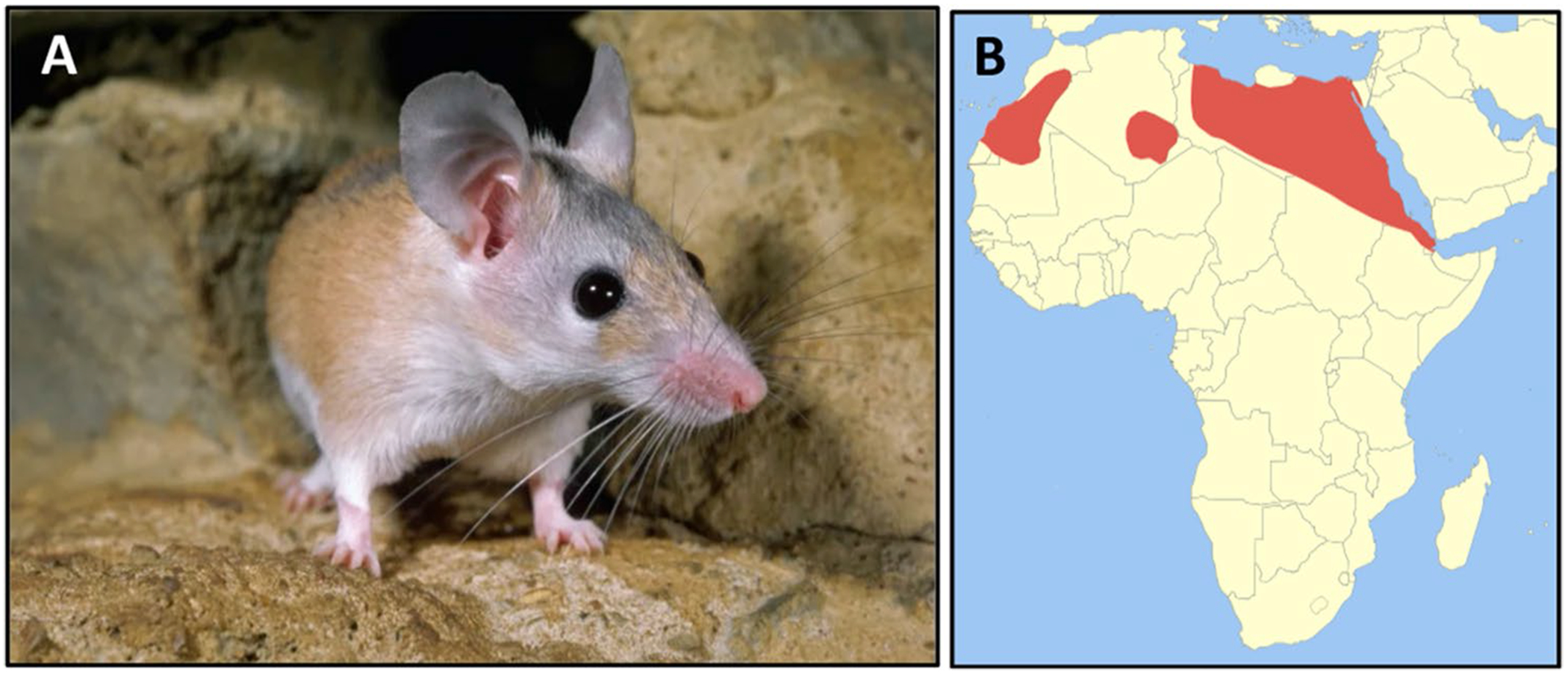
Fibrosis-driven solid organ failure is a major world-wide health burden with few therapeutic options. Spiny mice (genus: Acomys) are terrestrial mammals that regenerate severe skin wounds without fibrotic scars to evade predators. Recent studies have shown that spiny mice also regenerate acute ischemic and traumatic injuries to kidney, heart, spinal cord, and skeletal muscle. A common feature of this evolved wound healing response is a lack of formation of fibrotic scar tissue that degrades organ function, inhibits regeneration, and leads to organ failure. Complex tissue regeneration is an extremely rare property among mammalian species. In this article, we discuss the evidence that Acomys represents an emerging model organism that offers a unique opportunity for the biomedical community to investigate and clinically translate molecular mechanisms of scarless wound healing and regeneration of organ function in a mammalian species.

Keywords: Acomys; Evolution; Fibrosis; Wound healing.


Conflict of interest statement

Conflict of interest The authors declare that there are no financial or non-financial competing interests to disclose.

Figures

Fig. 1
Fig. 1
Acomys cahirinus—spiny mouse. A Adult spiny mice have narrow snouts, long whiskers, large ears, protruding black eyes, and spiny hairs along the dorsum. B These animals are adapted to hot arid climates and are found throughout northern Africa (red) and the Middle East (image licensed under the Creative Commons Attribution-Share Alike 3.0 Unported license-Wikimedia Commons)
Fig. 2
Fig. 2
Regeneration of ear hole injury in adult spiny mice. A Time course of ear hole closure and repigmentation in Mus (CD-1, top) and Acomys (spiny mouse, bottom). Note complete regeneration of ear hole injury in Acomys together with pigment and hair formation. B Histology (Safranin O stain). Left panel shows regeneration of normal ear architecture in vehicle-treated (mineral oil, MO) spiny mice with new cartilage (black dashed lines), adipocytes (asterisks), and hair follicles. (right panel) After treatment of Acomys with verteporfin (VP), an inhibitor of Yap-TEAD binding interactions (Brewer et al. 2021) between 3 and 5 weeks after ear punch injury, the resulting regenerated tissue is mispatterned and fibrotic. Significant reductions of new cartilage (dashed black lines) and adipocytes together with numerous ectopically located hair follicles appearing in the ventral region of de novo ear tissue at 5 weeks after injury (Brewer et al., 2021). Reprinted with permission from Developmental Cell
Fig. 3
Fig. 3
Regeneration of kidney function after acute renal injury in Acomys. (top left) Kidneys were injured either by permanent obstruction of the ureter (UUO, yellow arrow) or clamping of the renal artery for 40 min followed by release of the clamp allowing reperfusion of the ischemic kidney (IRI, blue arrow). (top right) Absence of fibrosis in obstructed kidneys by total collagen content measured as micrograms hydroxyproline per mg wet kidney weight in Acomys (red). By contrast, UUO produces a robust fibrotic response in MusB6 (blue) kidneys. (bottom left) Kidney function measured by blood urea nitrogen (BUN) levels 24 h after IRI injury. Note that both Acomys (Aco, red) and MusB6 (blue) exhibit similar levels of elevated BUN indicative of acute loss of kidney function. (bottom right) BUN levels at 2 weeks after IRI injury. Note further declines in kidney function in MusB6 (blue) progressing to organ failure. By contrast, BUN levels in Acomys (red) have returned to normal baseline values indicative of complete regeneration of organ function. Ns not significant, *p < 0.05, **p < 0.01, ***p < 0.001, **** p < 0.0001. (Okamura et al. 2021). Reprinted with permission from iScience. Figure created in BioRender
Fig. 4
Fig. 4
Acomys cahirinus (spiny mouse) (center top panel) is an emerging model organism to investigate and clinically translate molecular mechanisms of mammalian organ regeneration. Comparative studies have been reported of restored organ function after acute injury (from left to right) in kidney (Okamura et al. 2021); spinal cord (Nogueira-Rodrigues et al. 2022; Streeter et al. 2020), heart (Koopmans et al. 2021; Peng et al. 2021; Qi et al. 2021), skin (Brant et al. 2016; Brewer et al. 2021; Gawriluk et al. 2016; Harn et al. 2021; Matias et al. 2016; Jiang 2019; Seifert et al. 2012) and skeletal muscle (Maden et al. 2018). Some of the common findings across all studies of spiny mouse wound healing are indicated in the center boxed text. Figure created in BioRender

References

    1. Abbasi S, Sinha S, Labit E, Rosin NL, Yoon G, Rahmani W, Jaffer A, Sharma N, Hagner A, Shah P, Arora R, Yoon J, Islan A, Uchida A, Chang CK, Stratton JA, Scott RW, Rossi FMV, Underhill TM, Biernaskie J (2020) Distinct regulatory programs control the latent regenerative potential of dermal fibroblasts during wound healing. Cell Stem Cell 27:396–412. 10.1016/j.stem.2020.07.008 - DOI - PubMed
    1. Abrams MJ, Tan FH, Li Y, Basinger T, Heithe ML, Sarma A, Lee IT, Condiotte ZJ, Raffiee M, Dabiri JO, Gold DA, Goentoro L (2021) A conserved strategy for inducing appendage regeneration in moon jellyfish, Drosophila, and mice. eLife 10:e65092. 10.7554/eLife.65092 - DOI - PMC - PubMed
    1. Allan CH, Fleckman P, Fernandes RJ, Hager B, James J, Wisecarver Z, Satterstrom FK, Gutierrez A, Norman A, Pirrone A, Underwood RA, Rubin BP, Zhang M, Ramay HR, Clark JM (2006) Tissue response and Msx1 expression after human fetal digit tip amputation in vitro. Wound Repair Regen 14:398–404. 10.1111/j.1743-6109.2006.00139.x - DOI - PubMed
    1. Andres-Mateos E, Mejias R, Soleimani A, Lin BM, Burks TN, Marx R, Lin B, Zellars RC, Zhang Y, Huso DL, Marr TG, Leinwand LA, Merriman DK, Cohn RD (2012) Impaired skeletal muscle regeneration in the absence of fibrosis during hibernation in 13-lined ground squirrels. PLoS ONE 7:e48884. 10.1371/journal.pone.0048884 - DOI - PMC - PubMed
    1. Aurora AB, Porrello ER, Tan W, Mahmoud AI, Hill JA, Bassel-Duby R, Sadek HA, Olson EN (2014) Macrophages are required for neonatal heart regeneration. J Clin Invest 124:1382–1392. 10.1172/JCI72181 - DOI - PMC - PubMed

LinkOut - more resources