

Collagen fiber features and COL1A1: are they associated with elastic parameters in breast lesions, and can COL1A1 predict axillary lymph node metastasis?
- PMID: 36131254
- PMCID: PMC9490982
- DOI: 10.1186/s12885-022-10092-7
Collagen fiber features and COL1A1: are they associated with elastic parameters in breast lesions, and can COL1A1 predict axillary lymph node metastasis?
Abstract
Background: This study aimed to explore whether collagen fiber features and collagen type I alpha 1 (COL1A1) are related to the stiffness of breast lesions and whether COL1A1 can predict axillary lymph node metastasis (LNM).
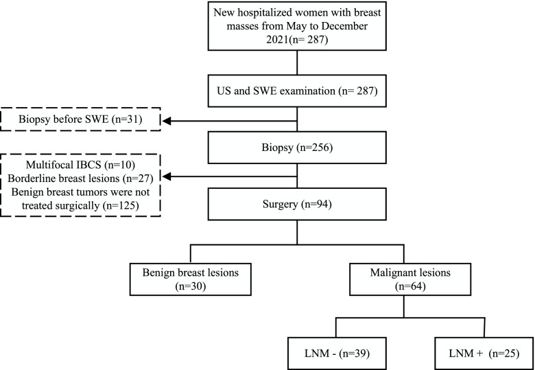
Methods: Ninety-four patients with breast lesions were consecutively enrolled in the study. Amongst the 94 lesions, 30 were benign, and 64 were malignant (25 were accompanied by axillary lymph node metastasis). Ultrasound (US) and shear wave elastography (SWE) were performed for each breast lesion before surgery. Sirius red and immunohistochemical staining were used to examine the shape and arrangement of collagen fibers and COL1A1 expression in the included tissue samples. We analyzed the correlation between the staining results and SWE parameters and investigated the effectiveness of COL1A1 expression levels in predicting axillary LNM.
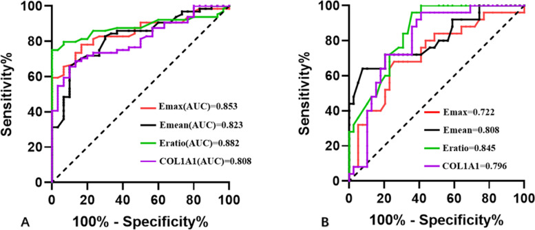
Results: The optimal cut-off values for Emax, Emean, and Eratio for diagnosing the benign and malignant groups, were 58.70 kPa, 52.50 kPa, and 3.05, respectively. The optimal cutoff for predicting axillary LNM were 107.5 kPa, 85.15 kPa, and 3.90, respectively. Herein, the collagen fiber shape and arrangement features in breast lesions were classified into three categories. One-way analysis of variance (ANOVA) showed that Emax, Emean, and Eratio differed between categories 0, 1, and 2 (P < 0.05). Meanwhile, elasticity parameters were positively correlated with collagen categories and COL1A1 expression. The COL1A1 expression level > 0.145 was considered the cut-off value, and its efficacy in benign and malignant breast lesions was 0.808, with a sensitivity of 66% and a specificity of 90%. Furthermore, when the COL1A1 expression level > 0.150 was considered the cut-off, its efficacy in predicting axillary LNM was 0.796, with sensitivity and specificity of 96% and 59%, respectively.
Conclusions: The collagen fiber features and expression levels of COL1A1 positively correlated with the elastic parameters of breast lesions. The expression of COL1A1 may help diagnose benign and malignant breast lesions and predict axillary LNM.
Keywords: Breast lesions; COL1A1; Collagen; Lymph node metastasis; SWE.
© 2022. The Author(s).
Conflict of interest statement
All authors disclosed no relevant relationships.
Figures


References
-
- Suvannarerg V, et al. Diagnostic performance of qualitative and quantitative shear wave elastography in differentiating malignant from benign breast masses, and association with the histological prognostic factors. Quant Imaging Med Surg. 2019;9(3):386–398. doi: 10.21037/qims.2019.03.04. - DOI - PMC - PubMed
MeSH terms
Substances
LinkOut - more resources
Full Text Sources
Medical
Miscellaneous
