

The surgical resection of dysplastic cerebellar gangliocytoma assisted by intraoperative sonography: illustrative case
- PMID: 36131570
- PMCID: PMC9563950
- DOI: 10.3171/CASE21451
The surgical resection of dysplastic cerebellar gangliocytoma assisted by intraoperative sonography: illustrative case
Abstract
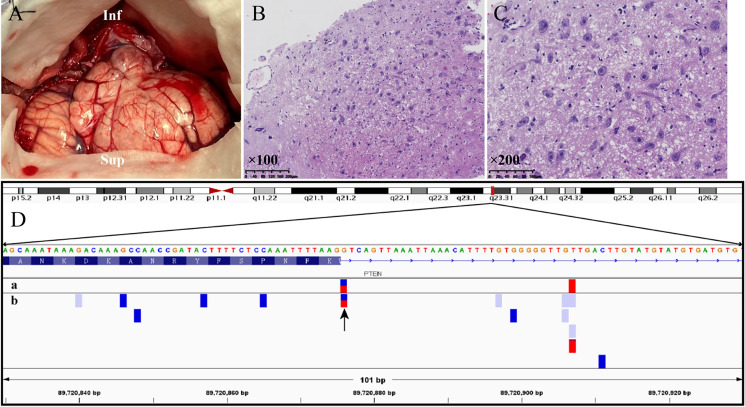
Background: Dysplastic gangliocytoma of the cerebellum (Lhermitte-Duclos disease) is an extremely rare, slow-growing hereditary mass lesion that is mainly characterized by both specific neuroradiological features and secondary hydrocephalus. Patients may present with symptoms of cerebellar mass lesion and increased intracranial pressure. As an important part of Cowden syndrome, Lhermitte-Duclos disease in adults is typically marked by PTEN gene mutation.
Observations: The clinical management of a 31-year-old woman who suffered Lhermitte-Duclos disease was introduced in this case report. Subtotal resection was performed with the assistance of intraoperative sonography to relieve obstructive hydrocephalus, and prophylactic C1 laminectomy was performed to prevent possible postoperative progression of the residual lesion. Perioperative care and surgical process were clearly revealed in an accompanying video. Intraoperative sonography of Lhermitte-Duclos disease presents hyperechoic distorted thickening cortices surrounded by hypoechoic edema belt. The patient did not report any significant neurological complications or sequelae after the lesion resection.
Lessons: The authors first reported the use of intraoperative sonography in resection of adult-onset Lhermitte-Duclos disease. Hopefully, the educative case report can provide a feasible experience in the diagnosis and treatment of Lhermitte-Duclos disease.
Keywords: Cowden syndrome; Lhermitte-Duclos disease; PTEN mutation; dysplastic gangliocytoma of the cerebellum; intraoperative sonography; surgical strategy.
Conflict of interest statement
Figures


Similar articles
-
Two illustrative cases of adult Lhermitte-Duclos disease and a systematic review of literature related to surgical management.Brain Spine. 2025 Apr 25;5:104258. doi: 10.1016/j.bas.2025.104258. eCollection 2025. Brain Spine. 2025. PMID: 40469941 Free PMC article.
-
Bilateral Recurrent Dysplastic Cerebellar Gangliocytoma (Lhermitte-Duclos Disease) in Cowden Syndrome: A Case Report and Literature Review.World Neurosurg. 2019 Jul;127:319-325. doi: 10.1016/j.wneu.2019.03.131. Epub 2019 Mar 21. World Neurosurg. 2019. PMID: 30905649 Review.
-
Radiographic Findings of Dysplastic Cerebellar Gangliocytoma (Lhermitte-Duclos Disease) in a Woman with Cowden Syndrome: A Case Study and Literature Review.J Radiol Case Rep. 2020 Mar 31;14(3):1-6. doi: 10.3941/jrcr.v14i3.3814. eCollection 2020 Mar. J Radiol Case Rep. 2020. PMID: 33082915 Free PMC article. Review.
-
Lhermitte-Duclos disease: A rare case of cerebellar tumor with successful surgical treatment.Surg Neurol Int. 2023 May 26;14:185. doi: 10.25259/SNI_302_2023. eCollection 2023. Surg Neurol Int. 2023. PMID: 37292412 Free PMC article.
-
Lhermitte-Duclos disease (dysplastic cerebellar gangliocytoma): a malformation, hamartoma or neoplasm?Acta Neurol Scand. 2002 Mar;105(3):137-45. doi: 10.1034/j.1600-0404.2002.1r127.x. Acta Neurol Scand. 2002. PMID: 11886354 Review.
Cited by
-
Lhermitte-Duclos disease in a 51-year old patient.Radiol Case Rep. 2024 Apr 24;19(7):2820-2825. doi: 10.1016/j.radcr.2024.03.057. eCollection 2024 Jul. Radiol Case Rep. 2024. PMID: 38689816 Free PMC article.
-
Lhermitte-Duclos disease: A series of six cases.J Neurosci Rural Pract. 2023 Jan-Mar;14(1):127-131. doi: 10.25259/JNRP-2022-3-10. Epub 2023 Jan 2. J Neurosci Rural Pract. 2023. PMID: 36891111 Free PMC article.
-
Two illustrative cases of adult Lhermitte-Duclos disease and a systematic review of literature related to surgical management.Brain Spine. 2025 Apr 25;5:104258. doi: 10.1016/j.bas.2025.104258. eCollection 2025. Brain Spine. 2025. PMID: 40469941 Free PMC article.
-
Utility of Intraoperative Ultrasound in Surgical Management of Lhermitte-Duclos Disease: A Case Report.Asian J Neurosurg. 2025 Mar 18;20(2):413-416. doi: 10.1055/s-0045-1805088. eCollection 2025 Jun. Asian J Neurosurg. 2025. PMID: 40485784 Free PMC article.
-
Treatment and Diagnostic Approach for Lhermitte-Duclos Disease and Suspected Cowden Syndrome.Cureus. 2024 Jun 23;16(6):e62968. doi: 10.7759/cureus.62968. eCollection 2024 Jun. Cureus. 2024. PMID: 39044874 Free PMC article.
References
-
- Louis DN, Perry A, Reifenberger G, et al. The 2016 World Health Organization classification of tumors of the central nervous system: a summary. Acta Neuropathol. 2016;131(6):803–820. - PubMed
-
- Lhermitte JDP. A diffuse cerebellar cortex ganglioneuroma. Bull Assoc Fr Etude Cancer. 1920;9:107.
-
- Khandpur U, Huntoon K, Smith-Cohn M, Shaw A, Elder JB. Bilateral recurrent dysplastic cerebellar gangliocytoma (Lhermitte-Duclos disease) in Cowden syndrome: a case report and literature review. World Neurosurg. 2019;127:319–325. - PubMed
-
- Nowak DA, Trost HA. Lhermitte-Duclos disease (dysplastic cerebellar gangliocytoma): a malformation, hamartoma or neoplasm? Acta Neurol Scand. 2002;105(3):137–145. - PubMed
-
- Koch R, Scholz M, Nelen MR, Schwechheimer K, Epplen JT, Harders AG. Lhermitte-Duclos disease as a component of Cowden’s syndrome. Case report and review of the literature. J Neurosurg. 1999;90(4):776–779. - PubMed
LinkOut - more resources
Full Text Sources
Research Materials
