. 2020 Jul 14;2(11):5192-5200.
doi: 10.1039/d0na00549e. eCollection 2020 Nov 11.

Neurite guidance and neuro-caging on steps and grooves in 2.5 dimensions

Affiliations

Neurite guidance and neuro-caging on steps and grooves in 2.5 dimensions

Cornelius Fendler et al. Nanoscale Adv. .

Abstract

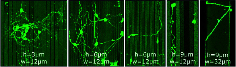
Directed guidance of neurites is a pre-requisite for tailor-made designs of interfaces between cells and semiconducting components. Grayscale lithography, reactive ion etching, and ultraviolet nanoimprint lithography are potent semiconductor industry-compatible techniques for a cost- and time-effective fabrication of modulated surfaces. In this work, neurite outgrowth of murine cerebellar neurons on 2.5D pathways produced with these methods is studied. Structures of micron-sized steps and grooves serve as cell culture platforms. The effects of contact guidance through topography and chemical guidance through selective poly-d-lysine coating on these platforms are analyzed. As a consequence, the herein presented fabrication approach can be utilized to cultivate and to study low-density neuronal networks in 2.5D configuration with a high degree of order.


Conflict of interest statement

There are no conflicts to declare.

Figures

Fig. 1
Fig. 1. (a–c) CLSM images of MCGCs cultured on ParC coated step structures produced by GSL at 4 DIV. Brightness of the steps correlates with the height: light and dark correspond to top steps and bottom pits, respectively. Red arrows point to neurites guided along edges. (d) Circular histogram of the normed distribution of neurite orientations in (a) relative to the step edges. (e–i) False-colored scanning electron microscopy (SEM) images of neurons on step-structures after 4 DIV.
Fig. 2
Fig. 2. CLSM images of MCGCs at 10–11 DIV on steps of varied height (h = 3 μm, 6 μm and 9 μm) and width (w = 12 μm and 32 μm) produced by RIE. All substrates have been coated with ParC and PDL.
Fig. 3
Fig. 3. False-colored SEM images and a CLSM image of MCGCs on chessboard structures produced by GSL after 6 DIV. (a) Two connected neurons. Three magnified areas of interest show (1) guidance along the bottom of a channel, (2) guidance on the sidewall of a channel, and (3) a neurite pathway exiting a channel. (b) Red arrows indicate spots, where neurites leave the channels. Yellow arrows indicate neurites, that are not guided. Blue arrows indicate re-entering of neurites into channels. Neurites originating from a cell cluster and somata that are settled on top of channels. (c–h) Examples of neurites exiting, entering, and crossing channels.
Fig. 4
Fig. 4. CLSM images and orientation analysis of neurons cultured on PDL circles with diameters of approx. 36 μm printed on an Al2O3 surface (7 DIV). (a–c) Circles printed in a pattern of 90 μm periodicity and an average distance between two circles of 54 μm. (d) Color-coded confocal image of neurons cultured on PDL circles in a pattern of 90 μm. Coloration of neurites corresponds to the neurite direction relative to the pattern. (e) Circular histogram of the normed distribution of neurite orientations on the pattern. (f) Percentage share of neurite pathways between nearest (first), second nearest and third-nearest circles. (g–i) Circles printed in a pattern of 190 μm and an average distance between two circles of 154 μm.
Fig. 5
Fig. 5. CLSM images of MCGCs at 6–8 DIV on chessboard structures produced by RIE and UV-NIL. (a) Cavities of 10 μm depth and steps in channels of 5 μm height coated with PDL with a materials printer. (b–e) Cavities of 14 μm depth and steps in channels of 4 μm height. PDL coated by a material printer (b) and with a micromanipulator (c–e). Red arrows indicate neurites guided at the top edge of cavities. Blue arrows indicate neurites at the bottom of cavities.

Similar articles

Cited by

  • Novel Concept of Micro Patterned Micro Titer Plates Fabricated via UV-NIL for Automated Neuronal Cell Assay Read-Out.
    Lohse M, Thesen MW, Haase A, Smolka M, Iceta NB, Ayerdi Izquierdo A, Ramos I, Salado C, Schleunitz A. Lohse M, et al. Nanomaterials (Basel). 2021 Apr 1;11(4):902. doi: 10.3390/nano11040902. Nanomaterials (Basel). 2021. PMID: 33916037 Free PMC article.
  • Robust neuronal differentiation of human iPSC-derived neural progenitor cells cultured on densely-spaced spiky silicon nanowire arrays.
    Harberts J, Siegmund M, Schnelle M, Zhang T, Lei Y, Yu L, Zierold R, Blick RH. Harberts J, et al. Sci Rep. 2021 Sep 22;11(1):18819. doi: 10.1038/s41598-021-97820-4. Sci Rep. 2021. PMID: 34552130 Free PMC article.
  • Interfacing with the Brain: How Nanotechnology Can Contribute.
    Ahmed AAA, Alegret N, Almeida B, Alvarez-Puebla R, Andrews AM, Ballerini L, Barrios-Capuchino JJ, Becker C, Blick RH, Bonakdar S, Chakraborty I, Chen X, Cheon J, Chilla G, Coelho Conceicao AL, Delehanty J, Dulle M, Efros AL, Epple M, Fedyk M, Feliu N, Feng M, Fernández-Chacón R, Fernandez-Cuesta I, Fertig N, Förster S, Garrido JA, George M, Guse AH, Hampp N, Harberts J, Han J, Heekeren HR, Hofmann UG, Holzapfel M, Hosseinkazemi H, Huang Y, Huber P, Hyeon T, Ingebrandt S, Ienca M, Iske A, Kang Y, Kasieczka G, Kim DH, Kostarelos K, Lee JH, Lin KW, Liu S, Liu X, Liu Y, Lohr C, Mailänder V, Maffongelli L, Megahed S, Mews A, Mutas M, Nack L, Nakatsuka N, Oertner TG, Offenhäusser A, Oheim M, Otange B, Otto F, Patrono E, Peng B, Picchiotti A, Pierini F, Pötter-Nerger M, Pozzi M, Pralle A, Prato M, Qi B, Ramos-Cabrer P, Genger UR, Ritter N, Rittner M, Roy S, Santoro F, Schuck NW, Schulz F, Şeker E, Skiba M, Sosniok M, Stephan H, Wang R, Wang T, Wegner KD, Weiss PS, Xu M, Yang C, Zargarian SS, Zeng Y, Zhou Y, Zhu D, Zierold R, Parak WJ. Ahmed AAA, et al. ACS Nano. 2025 Mar 25;19(11):10630-10717. doi: 10.1021/acsnano.4c10525. Epub 2025 Mar 10. ACS Nano. 2025. PMID: 40063703 Free PMC article. Review.
  • Nanomagnetic Guidance Shapes the Structure-Function Relationship of Developing Cortical Networks.
    Beck CL, Killeen CT, Johnson SC, Kunze A. Beck CL, et al. Nano Lett. 2024 Oct 30;24(43):13564-13573. doi: 10.1021/acs.nanolett.4c03156. Epub 2024 Oct 21. Nano Lett. 2024. PMID: 39432086

References

    1. Franze K. Janmey P. A. Guck J. Annu. Rev. Biomed. Eng. 2013;15:227–251. doi: 10.1146/annurev-bioeng-071811-150045. - DOI - PubMed
    1. Miller C. Jeftinija S. Mallapragada S. Tissue Eng. 2002;8:367–378. doi: 10.1089/107632702760184646. - DOI - PubMed
    1. Kleinfeld D. Kahler K. Hockberger P. J. Neurosci. 1988;8:4098–4120. doi: 10.1523/JNEUROSCI.08-11-04098.1988. - DOI - PMC - PubMed
    1. Oliva A. A. James C. D. Kingman C. E. Craighead H. G. Banker G. A. Neurochem. Res. 2003;28:1639–1648. doi: 10.1023/A:1026052820129. - DOI - PubMed
    1. Fan Y. W. Cui F. Z. Hou S. P. Xu Q. Y. Chen L. N. Lee I.-S. J. Neurosci. Methods. 2002;120:17–23. doi: 10.1016/S0165-0270(02)00181-4. - DOI - PubMed