

Dyke-Davidoff-Masson syndrome: Imaging diagnosis in an asymptomatic adult
- PMID: 36132061
- PMCID: PMC9483625
- DOI: 10.1016/j.radcr.2022.08.060
Dyke-Davidoff-Masson syndrome: Imaging diagnosis in an asymptomatic adult
Abstract
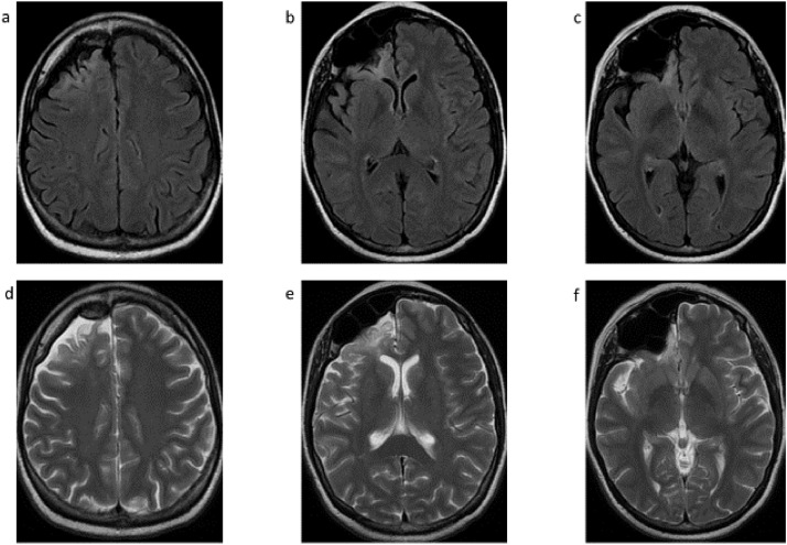
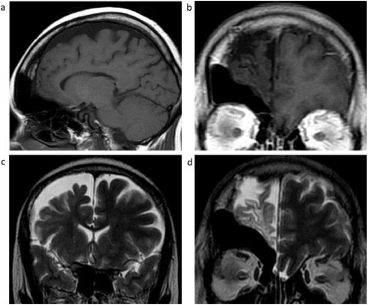
Dyke-Davidoff-Masson syndrome (DDMS) was first described in 1933 as a cerebral condition of hemispheric atrophy characterized clinically by contralateral hemiparesis, facial-asymmetry, seizures, and mental retardation. Neuroimaging findings include asymmetric thickening of the calvarium and enlargement of frontal and ethmoid sinuses. There have been 21 reported cases described in the literature with the syndrome undiagnosed until adult age, likely due to less severe or absent clinical findings or symptoms as described in the case presented in this report. This article describes a case where the Dyke-Davidoff-Masson imaging features were identified as an incidental finding on a CT scan of the brain performed for non-seizure related symptoms. A 54-year-old woman presented with weakness and gait difficulty and only upon further evaluation was she found to have cranial deformities. CT and MRI demonstrate encephalomalacia in the right frontal lobe anteriorly with gliosis and moderate unilateral cerebral atrophy, and extensive hypertrophy of the right frontal calvarium, right ethmoid cells and frontal sinuses.
Keywords: Calvarial Hypertrophy; Dyke-Davidoff-Masson syndrome; Neuroimaging findings.
© 2022 Published by Elsevier Inc. on behalf of University of Washington.
Figures

References
-
- Sharma S, Goyal D, Negi A, Sood RG, Jhobta A, Surya M. Dyke-Davidoff-Masson syndrome. Indian J Radiol Imaging. 2006;16:165–166.
-
- Stoevesandt D, Stock K, Spielmann RP, Heine HJ, Paulsen F, Bräuer L. Postmortal diagnosis of a Dyke-Davidoff-Masson syndrome in a 75-year-old woman: a case report. Ann Anat. 2009;191:225–227. - PubMed
-
- Atalar MH, Icagasioglu D, Tas F. Cerebral hemiatrophy (Dyke-Davidoff-Masson syndrome) in childhood: clinicoradiological analysis of 19 cases. Pediatr Int. 2007;49(1):70–75. - PubMed
Publication types
LinkOut - more resources
Full Text Sources
