The Expression of GPR30 in Iron-Overloaded Atypical Ovarian Epithelium and Ectopic Endometrium Is Closely Correlated with Transferrin Receptor and Pi3K
- PMID: 36132079
- PMCID: PMC9484882
- DOI: 10.1155/2022/8338874
The Expression of GPR30 in Iron-Overloaded Atypical Ovarian Epithelium and Ectopic Endometrium Is Closely Correlated with Transferrin Receptor and Pi3K
Abstract
Background: The mechanism of atypical hyperplasia of the ovarian epithelium and ectopic endometrium caused by iron overload remains unclear. Accordingly, we investigated possible effects on the human ovarian ectopic endometrium and ovarian epithelium by producing a high-iron environment with rat ovaries.
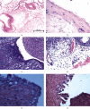
Objective: Human ovarian ectopic endometrium with atypical hyperplasia was collected, and the correlation between transferrin receptor GPR30 and Pi3K protein expression was studied by immunohistochemistry staining. Twenty SPF Sprague-Dawley female rats were microinjected with iron into one side of the ovary once a month, and the other ovary was used as the control. After 10 months of microinjection, the iron histological analysis was examined by Prussian blue staining, and ovarian endometrium morphology was assessed by HE staining. Abnormal lesion changes were measured by Pi3K staining. Evaluation of GPR30 was performed using reverse transcription PCR (RT-PCR) and western blotting, and the interrelationship between GPR30 and Pi3K was also assayed.
Results: GPR30 was significantly increased and correlated with the transferrin receptor and Pi3K in atypical human ovarian ectopic endometrium. Iron overload was confirmed in the 20 microinjected ovary cortexes, epithelial hyperplasia was observed in 12 ovaries, and papillary atypical hyperplasia was noted in eight ovaries. The RNA and protein levels of GPR30 were significantly increased in atypical hyperplasia compared to hyperplasia tissue samples. A positive relationship between GPR30 and Pi3K was found (P = 0.001).
Conclusion: The results suggest that persistent iron exposure may be a potential stimulus for ovarian endometriosis with atypical changes, and the abnormal increase in the new estrogen receptor GPR30 is closely related to this process.
Copyright © 2022 Li Long and Zhaoning Duan.
Conflict of interest statement
Authors Li Long and Zhao-ning Duan declare that they have no conflict of interest.
Figures


Similar articles
-
Transmembrane estrogen receptor GPR30 is more frequently expressed in malignant than benign ovarian endometriotic cysts and correlates with MMP-9 expression.Int J Gynecol Cancer. 2012 May;22(4):539-45. doi: 10.1097/IGC.0b013e318247323d. Int J Gynecol Cancer. 2012. PMID: 22495744
-
The expression status of G protein-coupled receptor GPR30 is associated with the clinical characteristics of endometriosis.Endocr Res. 2013;38(4):223-31. doi: 10.3109/07435800.2013.774011. Epub 2013 Mar 4. Endocr Res. 2013. PMID: 23458722
-
[Expression of G protein-coupled estrogen receptor and Gankyrin protein in ovarian endometriosis and its pathological significance].Zhong Nan Da Xue Xue Bao Yi Xue Ban. 2015 Aug;40(8):872-8. doi: 10.11817/j.issn.1672-7347.2015.08.008. Zhong Nan Da Xue Xue Bao Yi Xue Ban. 2015. PMID: 26333495 Chinese.
-
Ginsenoside Rg3 inhibits angiogenesis in a rat model of endometriosis through the VEGFR-2-mediated PI3K/Akt/mTOR signaling pathway.PLoS One. 2017 Nov 15;12(11):e0186520. doi: 10.1371/journal.pone.0186520. eCollection 2017. PLoS One. 2017. PMID: 29140979 Free PMC article.
-
Endometriosis: The Role of Iron Overload and Ferroptosis.Reprod Sci. 2020 Jul;27(7):1383-1390. doi: 10.1007/s43032-020-00164-z. Epub 2020 Feb 19. Reprod Sci. 2020. PMID: 32077077 Review.
Cited by
-
Future theranostic strategies: emerging ovarian cancer biomarkers to bridge the gap between diagnosis and treatment.Front Drug Deliv. 2024 Feb 1;4:1339936. doi: 10.3389/fddev.2024.1339936. eCollection 2024. Front Drug Deliv. 2024. PMID: 40836974 Free PMC article. Review.
References
-
- Guo D. H., Pang S. J., Shen Y. Atypical endometriosis: a clinicopathologic study of 163 cases. Zhonghua Fu Chan Ke Za Zhi . 2008;43(11):831–834. - PubMed
MeSH terms
Substances
LinkOut - more resources
Full Text Sources
Medical

