

Spontaneous self-assembly of amyloid β (1-40) into dimers
- PMID: 36132110
- PMCID: PMC9417245
- DOI: 10.1039/c9na00380k
Spontaneous self-assembly of amyloid β (1-40) into dimers
Abstract
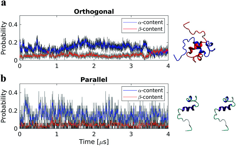
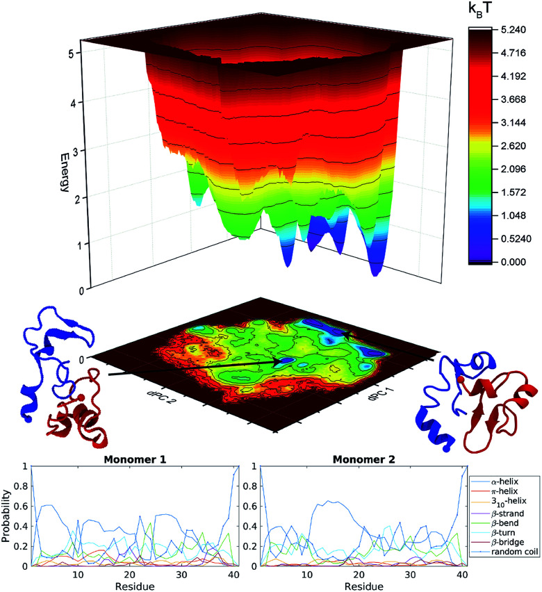
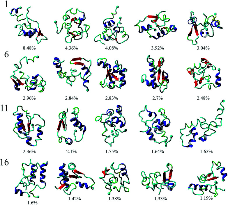
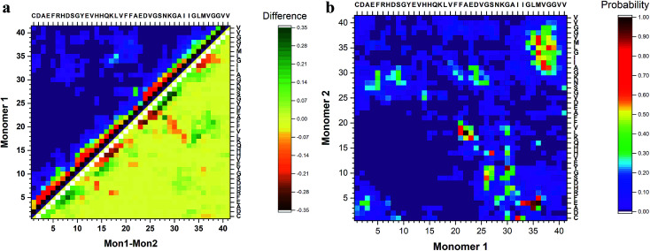
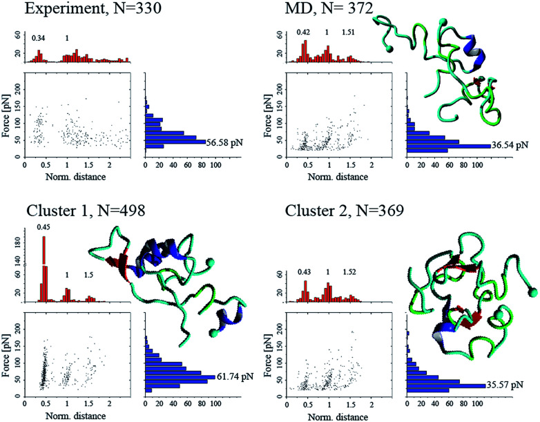
The self-assembly and fibrillation of amyloid β (Aβ) proteins is the neuropathological hallmark of Alzheimer's disease. However, the molecular mechanism of how disordered monomers assemble into aggregates remains largely unknown. In this work, we characterize the assembly of Aβ (1-40) monomers into dimers using long-time molecular dynamics simulations. Upon interaction, the monomers undergo conformational transitions, accompanied by change of the structure, leading to the formation of a stable dimer. The dimers are stabilized by interactions in the N-terminal region (residues 5-12), in the central hydrophobic region (residues 16-23), and in the C-terminal region (residues 30-40); with inter-peptide interactions focused around the N- and C-termini. The dimers do not contain long β-strands that are usually found in fibrils.
This journal is © The Royal Society of Chemistry.
Conflict of interest statement
Authors declare no competing interests.
Figures
Similar articles
-
Self-assembly of the full-length amyloid Aβ42 protein in dimers.Nanoscale. 2016 Dec 7;8(45):18928-18937. doi: 10.1039/c6nr06850b. Epub 2016 Oct 6. Nanoscale. 2016. PMID: 27714140 Free PMC article.
-
The inhibitory mechanism of a fullerene derivative against amyloid-β peptide aggregation: an atomistic simulation study.Phys Chem Chem Phys. 2016 May 14;18(18):12582-91. doi: 10.1039/c6cp01014h. Epub 2016 Apr 19. Phys Chem Chem Phys. 2016. PMID: 27091578
-
Norepinephrine Inhibits Alzheimer's Amyloid-β Peptide Aggregation and Destabilizes Amyloid-β Protofibrils: A Molecular Dynamics Simulation Study.ACS Chem Neurosci. 2019 Mar 20;10(3):1585-1594. doi: 10.1021/acschemneuro.8b00537. Epub 2019 Jan 15. ACS Chem Neurosci. 2019. PMID: 30605312
-
Elucidating the Structures of Amyloid Oligomers with Macrocyclic β-Hairpin Peptides: Insights into Alzheimer's Disease and Other Amyloid Diseases.Acc Chem Res. 2018 Mar 20;51(3):706-718. doi: 10.1021/acs.accounts.7b00554. Epub 2018 Mar 6. Acc Chem Res. 2018. PMID: 29508987 Free PMC article. Review.
-
Understanding amyloid fibril nucleation and aβ oligomer/drug interactions from computer simulations.Acc Chem Res. 2014 Feb 18;47(2):603-11. doi: 10.1021/ar4002075. Epub 2013 Dec 24. Acc Chem Res. 2014. PMID: 24368046 Review.
Cited by
-
On the Conformational Dynamics of β-Amyloid Forming Peptides: A Computational Perspective.Front Bioeng Biotechnol. 2020 Jun 3;8:532. doi: 10.3389/fbioe.2020.00532. eCollection 2020. Front Bioeng Biotechnol. 2020. PMID: 32656188 Free PMC article. Review.
-
Martini 3 coarse-grained model of enzymes: Framework with validation by all-atom simulations and x-ray diffraction measurements.J Chem Phys. 2025 Apr 7;162(13):135104. doi: 10.1063/5.0247634. J Chem Phys. 2025. PMID: 40177969
-
Amyloid Oligomers: A Joint Experimental/Computational Perspective on Alzheimer's Disease, Parkinson's Disease, Type II Diabetes, and Amyotrophic Lateral Sclerosis.Chem Rev. 2021 Feb 24;121(4):2545-2647. doi: 10.1021/acs.chemrev.0c01122. Epub 2021 Feb 5. Chem Rev. 2021. PMID: 33543942 Free PMC article. Review.
-
In-silico structural analysis of Heterocephalus glaber amyloid beta: an anti-Alzheimer's peptide.Mol Biol Res Commun. 2024;13(1):29-42. doi: 10.22099/mbrc.2023.48223.1862. Mol Biol Res Commun. 2024. PMID: 38164365 Free PMC article.
-
AFM Probing of Amyloid-Beta 42 Dimers and Trimers.Front Mol Biosci. 2020 Apr 24;7:69. doi: 10.3389/fmolb.2020.00069. eCollection 2020. Front Mol Biosci. 2020. PMID: 32391380 Free PMC article.
References
LinkOut - more resources
Full Text Sources