Review
. 2022 Sep 5:12:935231.
doi: 10.3389/fonc.2022.935231. eCollection 2022.

Proteolytic modulation of tumor microenvironment signals during cancer progression

Affiliations
Review

Proteolytic modulation of tumor microenvironment signals during cancer progression

Yoshifumi Itoh. Front Oncol. .

Abstract

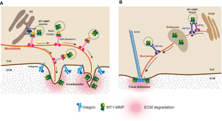
Under normal conditions, the cellular microenvironment is optimized for the proper functioning of the tissues and organs. Cells recognize and communicate with the surrounding cells and extracellular matrix to maintain homeostasis. When cancer arises, the cellular microenvironment is modified to optimize its malignant growth, evading the host immune system and finding ways to invade and metastasize to other organs. One means is a proteolytic modification of the microenvironment and the signaling molecules. It is now well accepted that cancer progression relies on not only the performance of cancer cells but also the surrounding microenvironment. This mini-review discusses the current understanding of the proteolytic modification of the microenvironment signals during cancer progression.

Keywords: ECM; Soluble factors; TME; invasion; matrikine; membrane protein shedding; proteinases.


Conflict of interest statement

The author declares that the research was conducted in the absence of any commercial or financial relationships that could be construed as a potential conflict of interest.

Figures

Figure 1
Figure 1
Proteolytic events during cancer progression. Epithelial cancer arises within the epithelial layer. They break through the basement membrane (BM) and invade stromal tissue. Upon BM degradation, matrikines are generated by proteolytic action, and cell surface extracellular matrix (ECM) receptor shedding promotes cancer cell motility. Stromal fibroblasts are activated and become cancer-associated fibroblasts and help cancer cells further invade. Tumor-associated macrophages (TAMs) help to evade the immune system. Cells degrade stromal the ECM further, including type I collagen, and intravasate into the vessel. Cancer cells traverse other organs through the bloodstream, interacting and rolling on the endothelial cell layer. Intercellular adhesion molecule–1 may be expressed in cancer cells, and its shedding allows cancer cells to migrate through the endothelial cell layer and extravasate. Cancer cells invade stromal tissue, form a metastatic colony, create a tumore microenvironment, and cause tissue malfunction.
Figure 2
Figure 2
Domain structure of metalloproteinases. Matrix metalloproteinases (MMPs) can be divided into two major groups: soluble MMPs and membrane-type MMPs. According to their structures, they can be classified into six soluble MMPs and two subgroups in membrane-type MMPs. MMP-11, 21, 28, 23, and MT-MMPs have a basic motif of RXKR that is recognized and cleaved by proprotein convertases to activate the enzymes by removing their pro-domain. Sig, signal peptide; Pro, pro-domain; Cat, catalytic domain; L, linker or hinge region; Hpx, hemopexin domain; C, cysteine; FN-II, fibronectin type-II repeats; TM, transmembrane domain; CA, cysteine array; IgG, IgG-like domain; L1, linker1 or hinge region; L2, linker 2 or stalk region; MT-Loop, eight amino acids insertion unique to TM-type MT-MMPs; and CP, cytoplasmic domain. ADAM enzymes have a conserved domain structure. DITG, a disintegrin-like domain; CysR, a cysteine-rich domain; EGF, an EGF-like domain. ADAMTS enzymes also have a conserved domain structure and differ in the number of thrombospondin motifs (TS) at their C-terminus. ADAMTS-4 is the smallest, without a C-terminal TS, and ADAMTS-5 and 8 have two. Other members have 2–14 repeats. Spacer, spacer domain. Both ADAM and ADAMTS enzymes have an RXKR motif at the C-terminus of their propeptide for activation by proprotein convertases.
Figure 3
Figure 3
Plasmin system. Plasminogen is a precursor form of plasmin, mainly produced in the liver and present in the plasma at approximately 150–200 μg/ml. Plasminogen is activated by either a tissue plasminogen activator (tPA) or urokinase plasminogen activator (uPA). uPA is bound to the glycosylphosphatidylinositol-anchored uPA receptor and activates plasminogen on the cell surface. tPA and uPA are also produced as precursor forms, and activated plasmin can activate these activators. Activated plasmin can degrade cross-linked fibrin and various ECM components, interact with the complement system to facilitate complement cascade, activate proMMPs, activate the precursor form of TGFβ, and cause syndecan shedding.
Figure 4
Figure 4
Vesicle transport of MT1-MMP to the invadopodia and the focal adhesion (FA). (A) Vesicle transport of MT1-MMP to the invadopodia has been extensively studied. It was shown that the endoplasmic reticulum protein protrudin plays a crucial role. Protrudin makes contact sites with RAB7 and phosphatidylinositol 3-phosphate (PI3P)–positive MT1-MMP-containing vesicles. Protrudin hands over RAB7-binding KIF5 adaptor protein FYCO1, enabling the transport of MT1-MMP-containing vesicles along microtubules toward invadopodia (32). (B) It was found that localization at FA is due to the direct transport of MT1-MMP-containing vesicles along the microtubules. KIF3A and KIF13A transport the vesicles between the trans-Golgi and the endosome. From the endosome, KIF13A solely transports the vesicles to the plasma membrane. The FA localization of MT1-MMP is essential for the HT1080 cell invasiveness (33).

References

    1. Hanahan D, Coussens LM. Accessories to the crime: functions of cells recruited to the tumor microenvironment. Cancer Cell (2012) 21(3):309–22. doi: 10.1016/j.ccr.2012.02.022 - DOI - PubMed
    1. Karamanos NK. Extracellular matrix: key structural and functional meshwork in health and disease. FEBS J (2019) 286(15):2826–9. doi: 10.1111/febs.14992 - DOI - PubMed
    1. Karamanos NK, Theocharis AD, Neill T, Iozzo RV. Matrix modeling and remodeling: A biological interplay regulating tissue homeostasis and diseases. Matrix Biol (2019) 75-76:1–11. doi: 10.1016/j.matbio.2018.08.007 - DOI - PMC - PubMed
    1. Piperigkou Z, Kyriakopoulou K, Koutsakis C, Mastronikolis S, Karamanos NK. Key matrix remodeling enzymes: Functions and targeting in cancer. Cancers (Basel) (2021) 13(6):1441. doi: 10.3390/cancers13061441 - DOI - PMC - PubMed
    1. Nagase H, Visse R, Murphy G. Structure and function of matrix metalloproteinases and TIMPs. Cardiovasc Res (2006) 69(3):562–73. doi: 10.1016/j.cardiores.2005.12.002 - DOI - PubMed

LinkOut - more resources