Review
. 2020 Jun 19;2(9):3632-3655.
doi: 10.1039/d0na00187b. eCollection 2020 Sep 16.

Cancer treatment by magneto-mechanical effect of particles, a review

Affiliations
Review

Cancer treatment by magneto-mechanical effect of particles, a review

Cécile Naud et al. Nanoscale Adv. .

Abstract

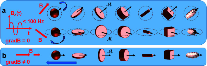
Cancer treatment by magneto-mechanical effect of particles (TMMEP) is a growing field of research. The principle of this technique is to apply a mechanical force on cancer cells in order to destroy them thanks to magnetic particles vibrations. For this purpose, magnetic particles are injected in the tumor or exposed to cancer cells and a low-frequency alternating magnetic field is applied. This therapeutic approach is quite new and a wide range of treatment parameters are explored to date, as described in the literature. This review explains the principle of the technique, summarizes the parameters used by the different groups and reports the main in vitro and in vivo results.


Conflict of interest statement

There are no conflicts to declare.

Figures

Fig. 1
Fig. 1. Sketch of various types of magnetic particles exposed to variable magnetic fields, thus subjected to magneto-mechanical torques (and/or forces), tending to rotate (and/or translate) the particles. Average magnetic moment of the particle: black arrows; applied magnetic field B: red arrows; particle rotation or translation direction: blue arrows. (a) Effect of the magnetic torque, generated by a spatially uniform magnetic field (gradB = 0), tending to align the magnetic moment of the particle with the direction of the magnetic field B, leading to the particle rotation. = (B). A rotating magnetic field can thus cause stable rotation or vibration of the particle, synchronized on its frequency (provided the frequency is low enough). (b) Magnetic force produced by the magnetic field gradient – derived from a spatially non-uniform magnetic field (i.e. gradB ≠ 0) – leading to the particle translation towards regions of large magnetic fields. Variable magnetic field gradient may also produce particle vibration.
Fig. 2
Fig. 2. Representative scheme showing properties of particles with different shapes, associated sizes and materials, and obtained results: microscopy images of particles and magnetization curves (magnetization as a function of magnetic field). Extracted from: (a and b) Shen et al., 2017 (ref. 30) with (a) magnetization curve of dry particles at 300 K and (b) TEM images of iron oxide particles doped with zinc (l = 62 nm) [Reproducted with permission (ref. 30), Copyright© 2017, Ivyspring International Publisher, Theranostics]. (c) Kilinc et al., 2015: SEM image of Fe–Au nanorods (d = 254 nm and l = 2 μm) [Reproducted with permission (ref. 34), Copyright© 2015, Wiley-VCH, Adv. Healthcare Mater.]. (d) Martínez et al., 2016: SEM image of Fe nanowire (l = 6.4 ± 1.3 μm and d = 30–40 nm) [Reproducted with permission (ref. 35), Copyright© 2016, Springer Nature, Sci. Rep.]. (e) Contreras et al., 2015: magnetization loops of an array of Ni nanowires (l = 4 μm and d = 30–40 nm) with magnetic field applied in the in-plane (black) and out-of-plane (red) directions [Reproducted with permission (ref. 33), Copyright© 2015, Dove Press, Int. J. Nanomed.]. (f and g) Wong et al., 2017 (ref. 36) with (f) hysteresis loop of NiFe particles with d = 150–350 nm (black to blue curve, respectively) and l = 500 nm, and (g) SEM images of NiFe particles of d = 350 nm and l = 75 nm, 200 nm and 500 nm (from left to right on the image) [Reproducted with permission (ref. 36), Copyright© 2017, Springer Nature, Sci. Rep.]. (h) Leulmi et al., 2015: SEM image of NiFe particles (d = 1.3 μm and l = 60 nm) [Reproducted with permission (ref. 38), Copyright© 2015, Royal Society of Chemistry, Nanoscale]. (i) Mansell et al., 2017: out-of-plane (red) and in-plane (black) hysteresis loops (b) for an array of 2 μm CoFeB/Pt particles and (d) for an array of 2 μm NiFe vortex particles [Reproducted with permission (ref. 27), Copyright© 2017, Springer Nature, Sci. Rep.]. (j) D. Cheng et al., 2014: TEM image of iron oxide particles (d = 200 ± 50 nm) [Reproducted with permission (ref. 32), Copyright© 2014, Springer, Nanoscale Res. Lett.]. (k) Wo et al., 2016: magnetization curve of hollow magnetic nanospheres of Fe3O4 (d = 250–550 nm) [Reproducted with permission (ref. 45), Copyright© 2016, Ivyspring International Publisher, Theranostics]. (l and m) Chiriac et al., 2018 (ref. 52) with (l) SEM image and (m) magnetization curve of Fe68.2Cr11.5Nb0.3B20 particles (l = 10–200 nm) [Reproducted with permission (ref. 52), Copyright© 2018, Springer Nature, Sci. Rep.].
Fig. 3
Fig. 3. Simplified diagrams of the mainly used magnetic field application devices. (a) Magnetic stirrer composed of two magnets at the end of a rotating rod (top view). (b) A coil powered by an alternating current that creates an alternating magnetic field inside or above the coil. The arrows in loops represent the magnetic flux line. (c) Ferrite core surrounded by a copper coil through which a sinusoidal alternating current flows. (d) System composed of 4 coils powered by an alternating sinusoidal current. The amplitude, phase shift and frequency of the applied current can be chosen to create an alternating or rotating field in the center of the 4 coils. (e) Halbach cylinder composed in this example of 8 permanent magnets creating an homogeneous field in the hollow of the cylinder. The rotating field is obtained by rotating the cylinder. (f) System composed of two magnets allowing to create a relatively homogeneous constant field.
Fig. 4
Fig. 4. (a) and (b) Extracted from Chiriac et al., 2018: human osteosarcoma cells (a) before and (b) after the magneto-mechanical actuation (rotating field). Live cells are colored in green and dead cells in red [Reproducted with permission (ref. 52), Copyright© 2018, Springer Nature, Sci. Rep.]; (c–f) extracted from Hu and Gao, 2010: prostate cancer cells after treatment: (c) cells only, (d) cells exposed to magnetic field, (e) cells with particles, (f) cells with particles and exposed to magnetic field. Particles are biphasic iron oxide nanocomposites (d = 180 nm). Rotating magnetic field (0.83 Hz) was applied for 15 min. Dead cells appear blue due to trypan blue staining [Reproducted with permission (ref. 41), Copyright© 2010, American Chemical Society, J. Am. Chem. Soc.]; (g and h) extracted from Liu et al., 2012: cell membrane topographical imaging by AFM. (g) Control group. Surface of untreated cell was smooth. (h) MCF-7 cell treated by multiwalled carbon nanotubes exposed in 40 mT magnetic field for 20 min. Surface of the treated group is much rougher than controls with many small pore like structures [Reproducted with permission (ref. 31), Copyright© 2012, American Chemical Society, Nano Lett.].
Fig. 5
Fig. 5. Extracted from Y. Cheng et al., 2015:in vivo therapeutic efficacy of the magnetic particles (MPs) under rotating magnetic field. “The U87 cells were pre-incubated with MPs for 24 h and implanted in the mouse brain”. (a) Quantification of the tumor bioluminescence signal over 4 weeks (n = 5 mice per group). Data are presented as mean ± SE. **p < 0.01, ***p < 0.001 (Student's t test); (b) Kaplan–Meier survival curve of the mice with and without magnetic field treatment. *p < 0.05 (log rank test) [Reproducted with permission (ref. 37), Copyright© 2015, Elsevier B.V., J. Control. Release].

References

    1. Pankhurst Q. A. Connolly J. Jones S. K. Dobson J. P. Applications of magnetic nanoparticles in biomedicine. J. Phys. D: Appl. Phys. 2003;36:167–181. doi: 10.1088/0022-3727/36/13/201. - DOI
    1. Cheng Y. Morshed R. A. Auffinger B. Tobias A. L. Lesniak M. S. Multifunctional nanoparticles for brain tumor imaging and therapy. Adv. Drug Delivery Rev. 2014;66:42–57. doi: 10.1016/j.addr.2013.09.006. - DOI - PMC - PubMed
    1. Stephen Z. R. Kievit F. M. Zhang M. Magnetite nanoparticles for medical MR imaging. Mater. Today. 2011;14:330–338. doi: 10.1016/S1369-7021(11)70163-8. - DOI - PMC - PubMed
    1. Jin R. Lin B. Li D. Ai H. Superparamagnetic iron oxide nanoparticles for MR imaging and therapy: design considerations and clinical applications. Curr. Opin. Pharmacol. 2014;18:18–27. doi: 10.1016/j.coph.2014.08.002. - DOI - PubMed
    1. Arruebo M. Fernández-pacheco R. Ibarra M. R. Santamaría J. Magnetic nanoparticles for drug delivery. Nano Today. 2007;2:22–32. doi: 10.1016/S1748-0132(07)70084-1. - DOI