. 2021 May 17;3(13):3824-3834.
doi: 10.1039/d1na00211b. eCollection 2021 Jun 30.

Optimal centrifugal isolating of liposome-protein complexes from human plasma

Affiliations

Optimal centrifugal isolating of liposome-protein complexes from human plasma

Luca Digiacomo et al. Nanoscale Adv. .

Abstract

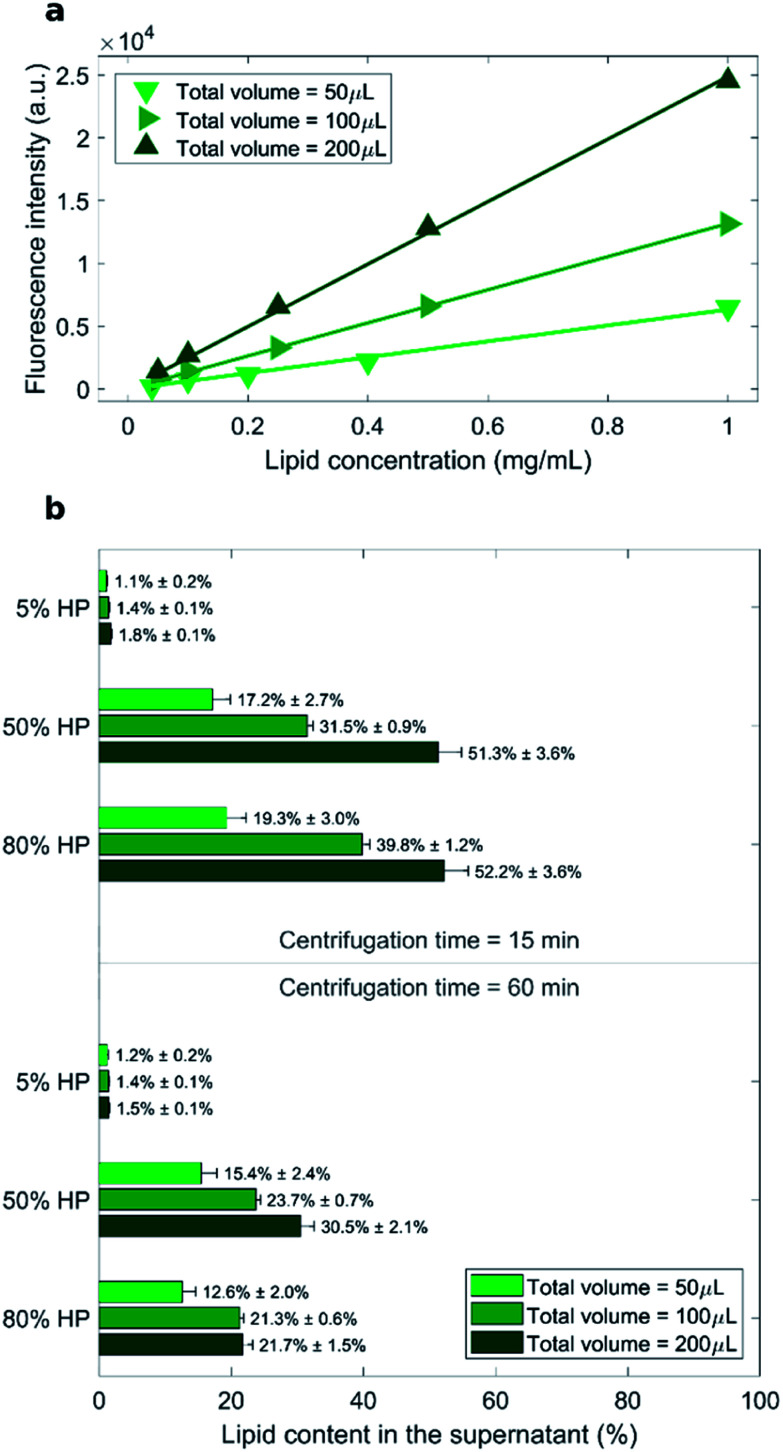
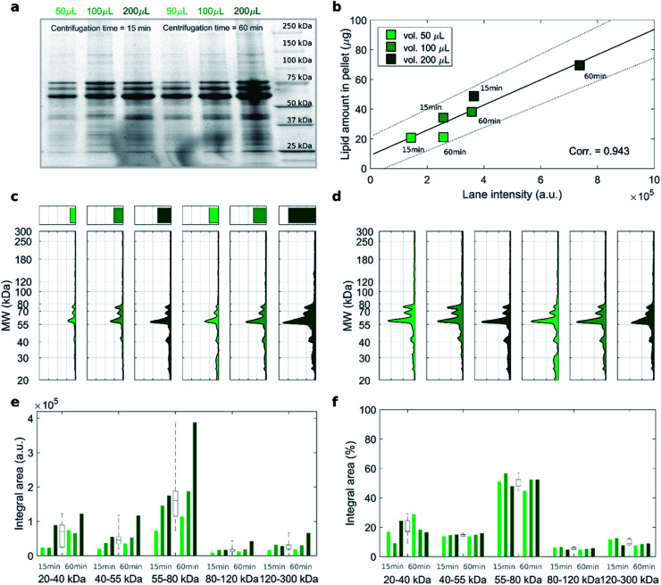
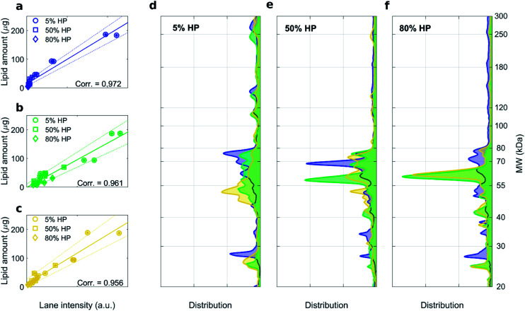
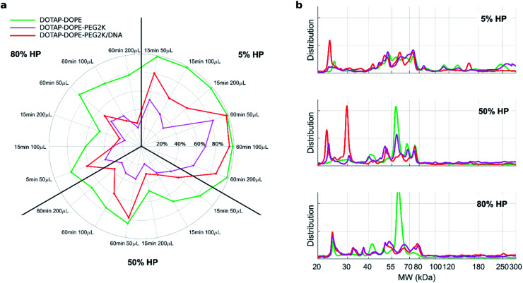
In the past few years, characterization of the protein corona (PC) that forms around liposomal systems has gained increasing interest for the development of novel therapeutic and diagnostic technologies. At the crossroads of fast-moving research fields, the interdisciplinarity of protein corona investigations poses challenges for experimental design and reporting. Isolation of liposome-protein complexes from biological fluids has been identified as a fundamental step of the entire workflow of PC characterization but exact specifications for conditions to optimize pelleting remain elusive. In the present work, key factors affecting precipitation of liposome-protein complexes by centrifugation, including time of centrifugation, total sample volume, lipid : protein ratio and contamination from biological NPs were comprehensively evaluated. Here we show that the total amount of isolated liposome-protein complexes and the extent of contamination from biological NPs may vary with influence factors. Our results provide protein corona researchers with precise indications to separate liposome-protein complexes from protein-rich fluids and include proper controls, thus they are anticipated to catalyze improved consistency of data mining and computational modelling of protein corona composition.


Conflict of interest statement

There are no conflicts to declare.

Figures

Fig. 1
Fig. 1. (a) Calibration curves for the measurements of lipid concentration by fluorescence experiments. (b) Measured recovery rates of lipids in the supernatant represented by percentage ratio of the lipid amount after precipitation to its initial amount (before precipitation).
Fig. 2
Fig. 2. (a) Cropped 1D SDS–PAGE image of the PC isolated from DOTAP–DOPE 1 : 1 (mol mol−1) after incubation with HP (50% vol) and centrifugation under the indicated conditions of sample volume and centrifugation time. (b) Correlation analysis between total lane intensity from 1D SDS–PAGE and recovered lipid amount in pellets, measured by fluorescence experiments. (c) Total lane intensity (bar) and absolute intensity profiles for each lane. (d) Corresponding normalized profiles. Integral areas within the specified molecular weight ranges for (e) absolute and (f) normalized profiles. For each MW range, integral area distributions are reported as grey boxplots. Despite huge differences in the absolute integral areas among the explored centrifugation conditions, the corresponding analysis on normalized profiles reveals no significant differences, thus indicates that the PC composition did not depend on centrifugation factors.
Fig. 3
Fig. 3. 1D SDS–PAGE analysis of the PC isolated from DOTAP–DOPE 1 : 3 (mol mol−1) (blue), DOTAP–DOPE 1 : 1 (mol mol−1) (green), DOTAP–DOPE 3 : 1 (mol mol−1) (yellow) under different centrifugation conditions (gel images are provided in Fig. S3, S1 and S4 in the ESI, respectively). (a–c) Correlation analysis between total lane intensity from 1D SDS–PAGE and recovered lipid amount in pellets, measured by fluorescence experiments. (d–f) Average normalized 1D SDS–PAGE profiles grouped by HP concentration.
Fig. 4
Fig. 4. (a) Polar plots of recovery rates of lipids in the pellets for unPEGylated DOTAP:DOPE liposomes, PEGylated liposome and PEGylated lipoplex. The percentage ratio of the lipid amount after precipitation to its initial amount (before precipitation) is represented by the distance from the center, for each of the investigated centrifugation conditions (radii of the polar plot). (b) 1D SDS–PAGE normalized profiles of the PC isolated from unPEGylated liposome, PEGylated liposome and PEGylated lipoplex at different HP concentration: 5% HP, 50% HP, 80% HP. For each experimental condition DOTAP and DOPE were mixed in equimolar ratio (i.e. 1 : 1 mol mol−1).
Fig. 5
Fig. 5. Nano liquid chromatography tandem mass spectrometry (nano LC-MS/MS) experiments. Volcano plots depicting the RPA-ratio between the two explored centrifugation conditions (i.e. total volume 50 μL, 60 min centrifugation time and total volume 200 μL and 15 min centrifugation time) as logarithmic fold change, vs. the corresponding p-value from Student's t test, for (a) DOTAP:DOPE liposomes, (b) PEGylated liposome and (c) PEGylated lipoplex. Proteins exhibiting relevant differences (p < 0.05 and |log2(fold change)| > 1) are indicated as colored dots, their cumulative RPA and total number are reported within the corresponding outer regions. Cumulative RPA and total number of proteins with similar abundances between the explored conditions (i.e. p ≥ 0.05 or |log2(fold change)| ≤ 1, grey dots) are reported in the central region.
Fig. 6
Fig. 6. (a) 1D SDS–PAGE image of PC for human plasma (HP) and liposomal formulations upon incubation with HP (50% vol), total volume = 50 μL and centrifugation time = 60 min. (b) Total lane intensity for each lane. (c–g) Corresponding absolute intensity profiles for the liposomal formulations, whereas the absolute profile of HP is superimposed as black shaded area.

Comment in

Similar articles

Cited by

References

    1. Liu J. Zhang R. Xu Z. P. Small. 2019:1900262. doi: 10.1002/smll.201900262. - DOI - PubMed
    1. Walkey C. D. Chan W. C. W. Chem. Soc. Rev. 2012;41:2780–2799. doi: 10.1039/C1CS15233E. - DOI - PubMed
    1. Caracciolo G. Farokhzad O. C. Mahmoudi M. Trends Biotechnol. 2017;35:257–264. doi: 10.1016/j.tibtech.2016.08.011. - DOI - PubMed
    1. Cedervall T. Lynch I. Lindman S. Berggård T. Thulin E. Nilsson H. Dawson K. A. Linse S. Proc. Natl. Acad. Sci. U. S. A. 2007;104:2050–2055. doi: 10.1073/pnas.0608582104. - DOI - PMC - PubMed
    1. Lundqvist M. Stigler J. Elia G. Lynch I. Cedervall T. Dawson K. A. Proc. Natl. Acad. Sci. U. S. A. 2008;105:14265–14270. doi: 10.1073/pnas.0805135105. - DOI - PMC - PubMed