

Therapeutic response differences between 2D and 3D tumor models of magnetic hyperthermia
- PMID: 36133021
- PMCID: PMC9418625
- DOI: 10.1039/d1na00224d
Therapeutic response differences between 2D and 3D tumor models of magnetic hyperthermia
Abstract
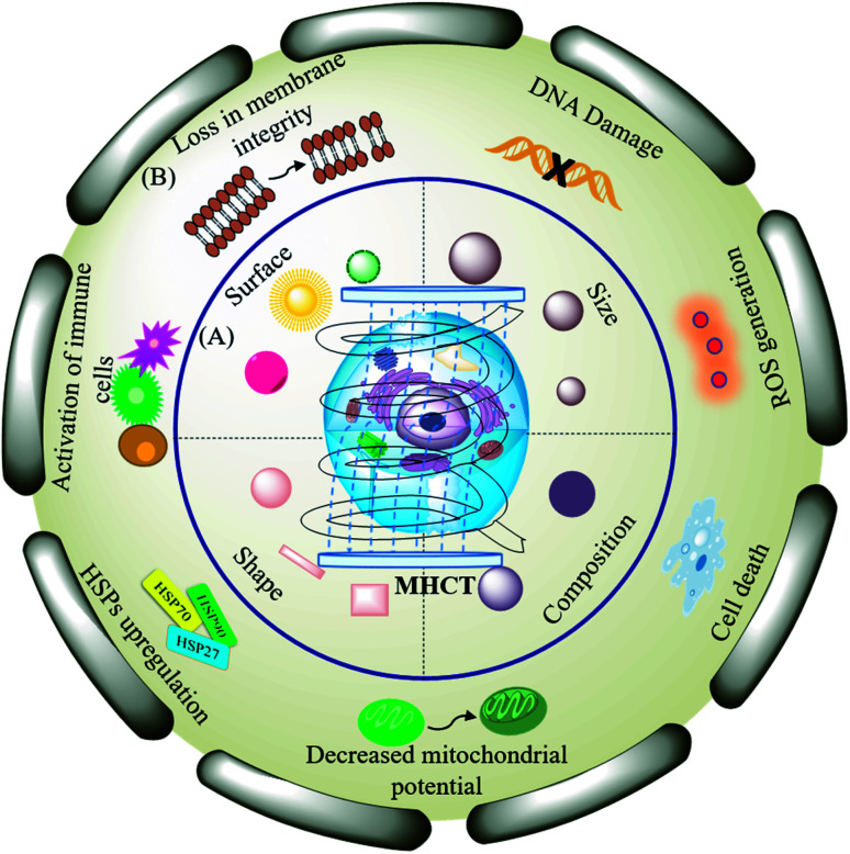
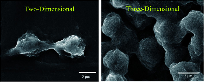
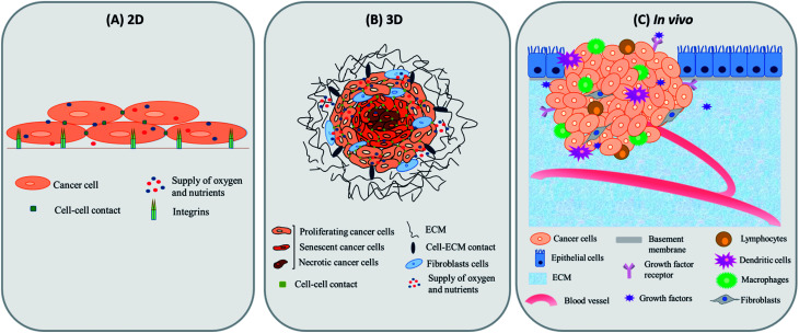
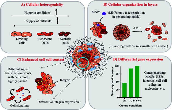
Magnetic hyperthermia-based cancer therapy (MHCT) has surfaced as one of the promising techniques for inaccessible solid tumors. It involves generation of localized heat in the tumor tissues on application of an alternating magnetic field in the presence of magnetic nanoparticles (MNPs). Unfortunately, lack of precise temperature and adequate MNP distribution at the tumor site under in vivo conditions has limited its application in the biomedical field. Evaluation of in vitro tumor models is an alternative for in vivo models. However, generally used in vitro two-dimensional (2D) models cannot mimic all the characteristics of a patient's tumor and hence, fail to establish or address the experimental variables and concerns. Considering that three-dimensional (3D) models have emerged as the best possible state to replicate the in vivo conditions successfully in the laboratory for most cell types, it is possible to conduct MHCT studies with higher clinical relevance for the analysis of the selection of magnetic parameters, MNP distribution, heat dissipation, action and acquired thermotolerance in cancer cells. In this review, various forms of 3D cultures have been considered and the successful implication of MHCT on them has been summarized, which includes tumor spheroids, and cultures grown in scaffolds, cell culture inserts and microfluidic devices. This review aims to summarize the contrast between 2D and 3D in vitro tumor models for pre-clinical MHCT studies. Furthermore, we have collated and discussed the usefulness, suitability, pros and cons of these tumor models. Even though numerous cell culture models have been established, further investigations on the new pre-clinical models and selection of best fit model for successful MHCT applications are still necessary to confer a better understanding for researchers.
This journal is © The Royal Society of Chemistry.
Conflict of interest statement
There are no conflicts to declare.
Figures


Similar articles
-
Evolution of Magnetic Hyperthermia for Glioblastoma Multiforme Therapy.ACS Chem Neurosci. 2019 Mar 20;10(3):1157-1172. doi: 10.1021/acschemneuro.8b00652. Epub 2019 Feb 19. ACS Chem Neurosci. 2019. PMID: 30715851 Review.
-
Expansion of thermometry in magnetic hyperthermia cancer therapy: antecedence and aftermath.Nanomedicine (Lond). 2022 Sep;17(21):1607-1623. doi: 10.2217/nnm-2022-0095. Epub 2022 Nov 1. Nanomedicine (Lond). 2022. PMID: 36318111 Review.
-
Functionalized manganese iron oxide nanoparticles: a dual potential magneto-chemotherapeutic cargo in a 3D breast cancer model.Nanoscale. 2023 Oct 5;15(38):15686-15699. doi: 10.1039/d3nr02816j. Nanoscale. 2023. PMID: 37724853
-
Optimization Study on Specific Loss Power in Superparamagnetic Hyperthermia with Magnetite Nanoparticles for High Efficiency in Alternative Cancer Therapy.Nanomaterials (Basel). 2020 Dec 26;11(1):40. doi: 10.3390/nano11010040. Nanomaterials (Basel). 2020. PMID: 33375292 Free PMC article.
-
In Silico Approach to Model Heat Distribution of Magnetic Hyperthermia in the Tumoral and Healthy Vascular Network Using Tumor-on-a-Chip to Evaluate Effective Therapy.Pharmaceutics. 2024 Aug 31;16(9):1156. doi: 10.3390/pharmaceutics16091156. Pharmaceutics. 2024. PMID: 39339193 Free PMC article.
Cited by
-
Co-Formulation of Iron Oxide and PLGA Nanoparticles to Deliver Curcumin and IFNα for Synergistic Anticancer Activity in A375 Melanoma Skin Cancer Cells.Pharmaceutics. 2025 Jun 30;17(7):860. doi: 10.3390/pharmaceutics17070860. Pharmaceutics. 2025. PMID: 40733069 Free PMC article.
-
Advances in screening hyperthermic nanomedicines in 3D tumor models.Nanoscale Horiz. 2024 Feb 26;9(3):334-364. doi: 10.1039/d3nh00305a. Nanoscale Horiz. 2024. PMID: 38204336 Free PMC article. Review.
-
Comparative heating efficiency and cytotoxicity of magnetic silica nanoparticles for magnetic hyperthermia treatment on human breast cancer cells.3 Biotech. 2022 Nov;12(11):313. doi: 10.1007/s13205-022-03377-y. Epub 2022 Oct 8. 3 Biotech. 2022. PMID: 36276464 Free PMC article.
-
Leveraging Xenobiotic-Responsive Cancer Stemness in Cell Line-Based Tumoroids for Evaluating Chemoresistance: A Proof-of-Concept Study on Environmental Susceptibility.Int J Mol Sci. 2024 Oct 23;25(21):11383. doi: 10.3390/ijms252111383. Int J Mol Sci. 2024. PMID: 39518936 Free PMC article.
-
FSP1 is a predictive biomarker of osteosarcoma cells' susceptibility to ferroptotic cell death and a potential therapeutic target.Cell Death Discov. 2024 Feb 17;10(1):87. doi: 10.1038/s41420-024-01854-2. Cell Death Discov. 2024. PMID: 38368399 Free PMC article.
References
-
- Datta N. R. Krishnan S. Speiser D. E. Neufeld E. Kuster N. Bodis S. Hofmann H. Magnetic nanoparticle-induced hyperthermia with appropriate payloads: Paul Ehrlich's “magic (nano)bullet” for cancer theranostics? Cancer Treat. Rev. 2016;50:217–227. doi: 10.1016/j.ctrv.2016.09.016. doi: 10.1016/j.ctrv.2016.09.016. - DOI - DOI - PubMed
-
- Liu X. Zhang Y. Wang Y. Zhu W. Li G. Ma X. Zhang Y. Chen S. Tiwari S. Shi K. Zhang S. Fan H. M. Zhao Y. X. Liang X. J. Comprehensive understanding of magnetic hyperthermia for improving antitumor therapeutic efficacy. Theranostics. 2020;10:3793–3815. doi: 10.7150/thno.40805. doi: 10.7150/thno.40805. - DOI - DOI - PMC - PubMed
Publication types
LinkOut - more resources
Full Text Sources
Miscellaneous