. 2021 Jan 4;3(4):1029-1046.
doi: 10.1039/d0na00820f. eCollection 2021 Feb 23.

Tunable magnetothermal properties of cobalt-doped magnetite-carboxymethylcellulose ferrofluids: smart nanoplatforms for potential magnetic hyperthermia applications in cancer therapy

Affiliations

Tunable magnetothermal properties of cobalt-doped magnetite-carboxymethylcellulose ferrofluids: smart nanoplatforms for potential magnetic hyperthermia applications in cancer therapy

Alice G Leonel et al. Nanoscale Adv. .

Abstract

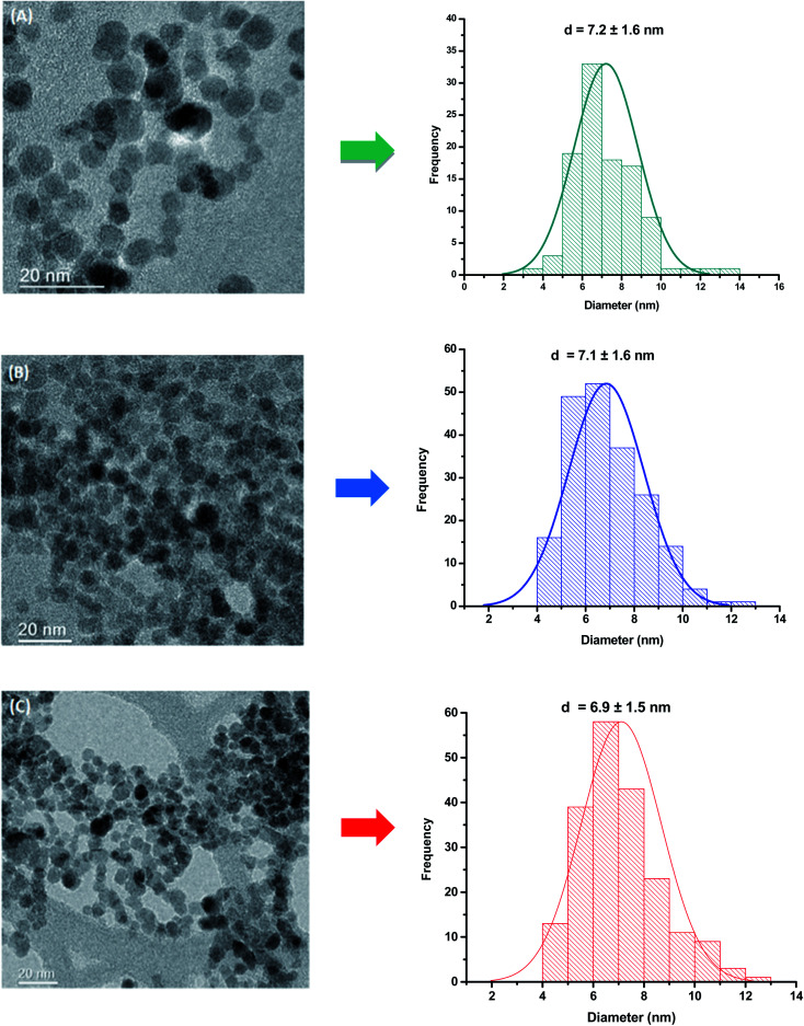
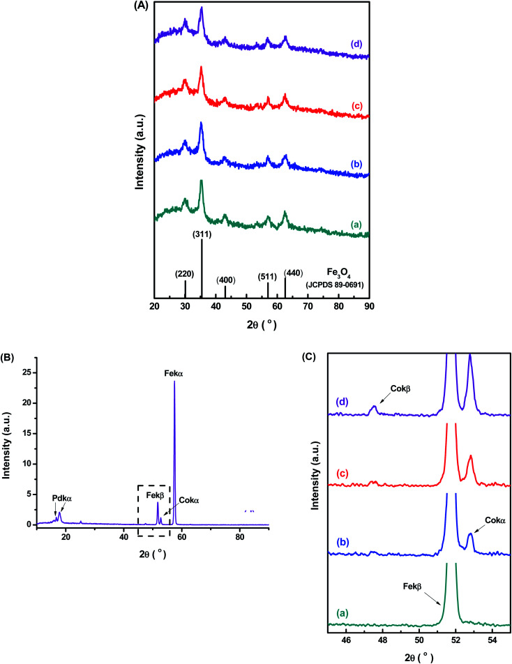
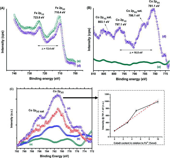
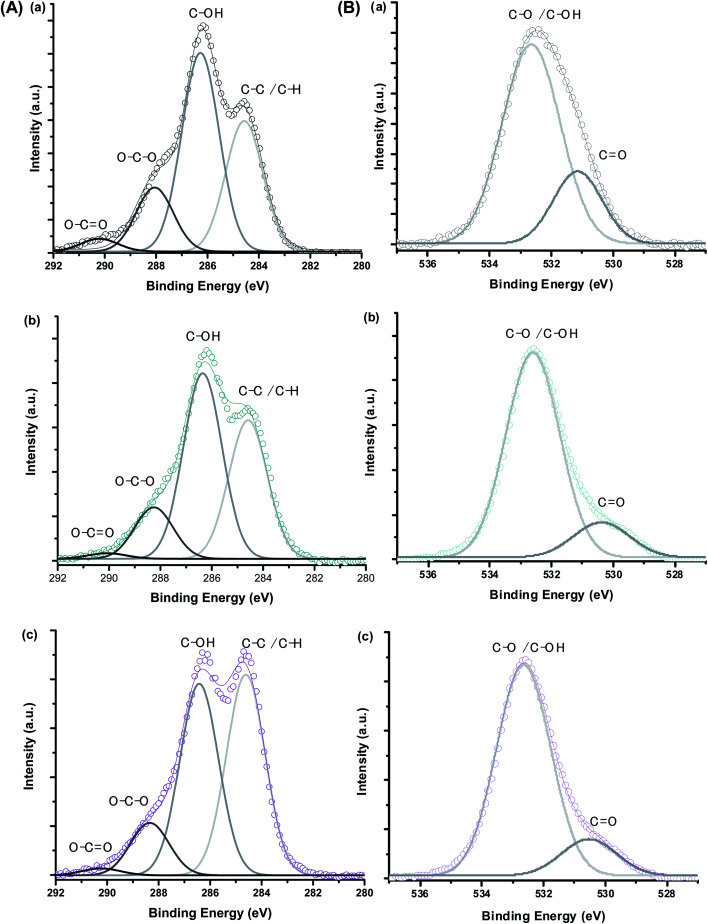
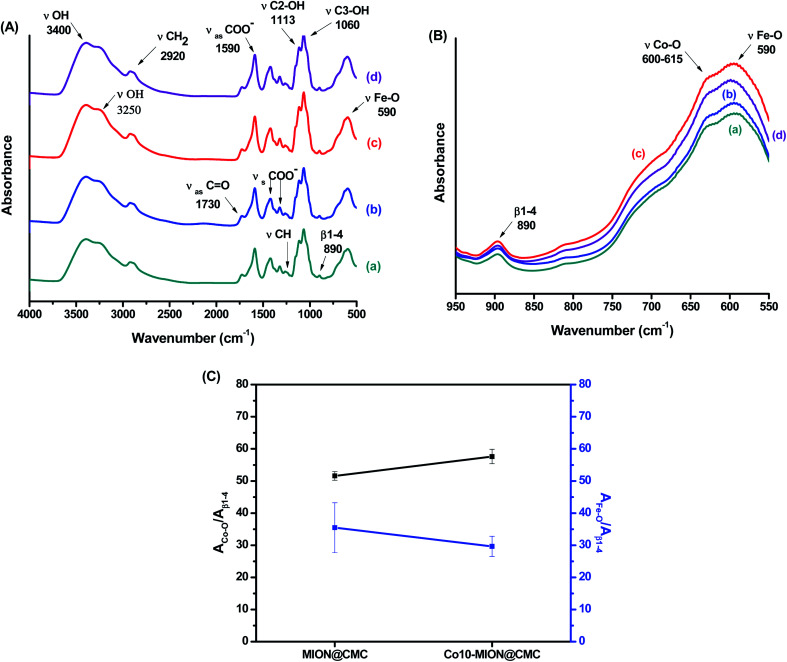
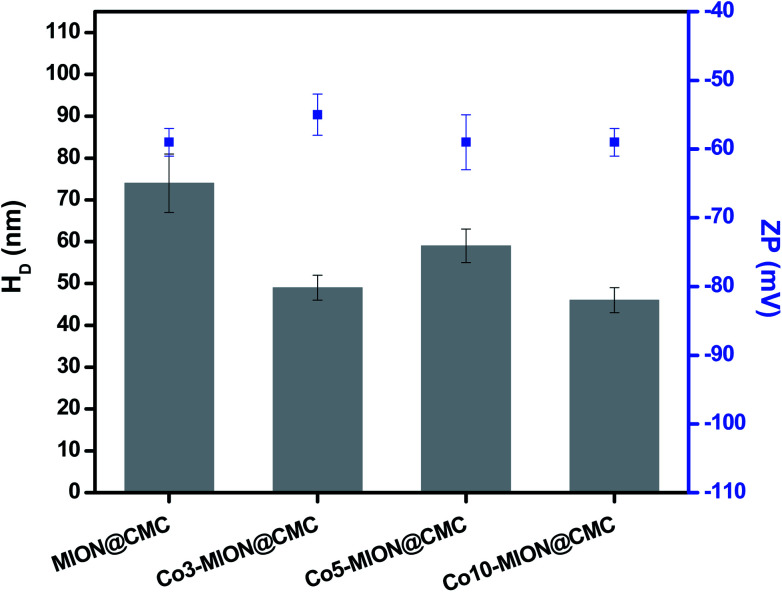
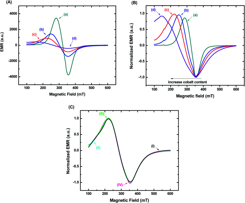
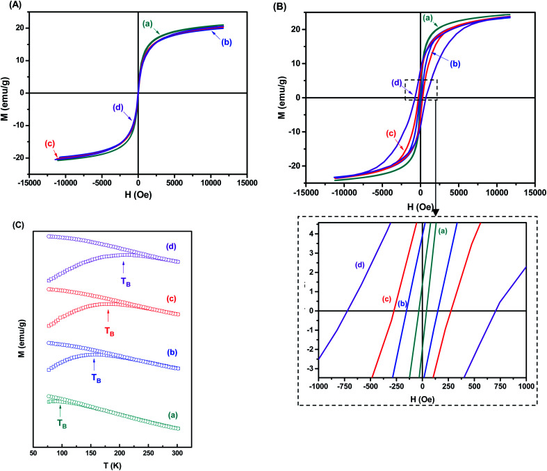
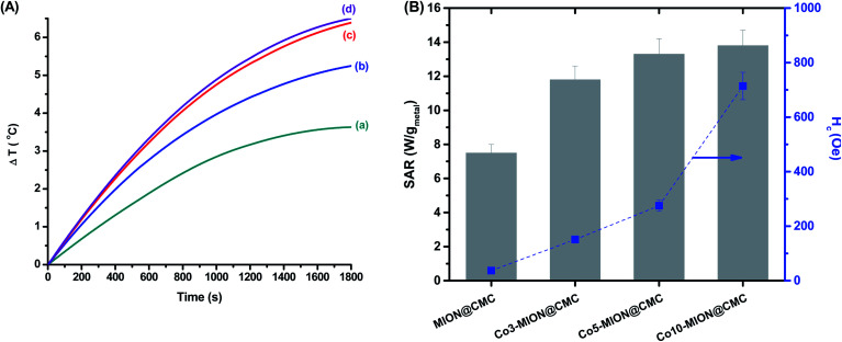
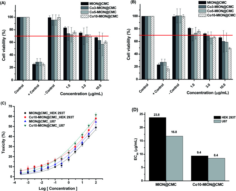
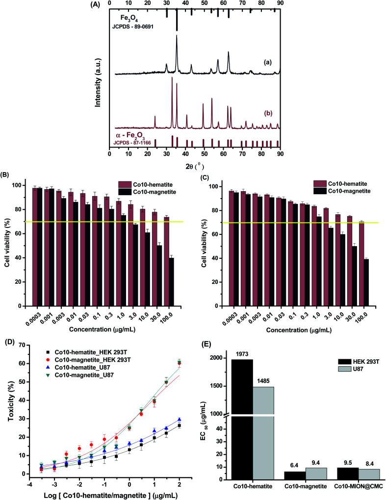
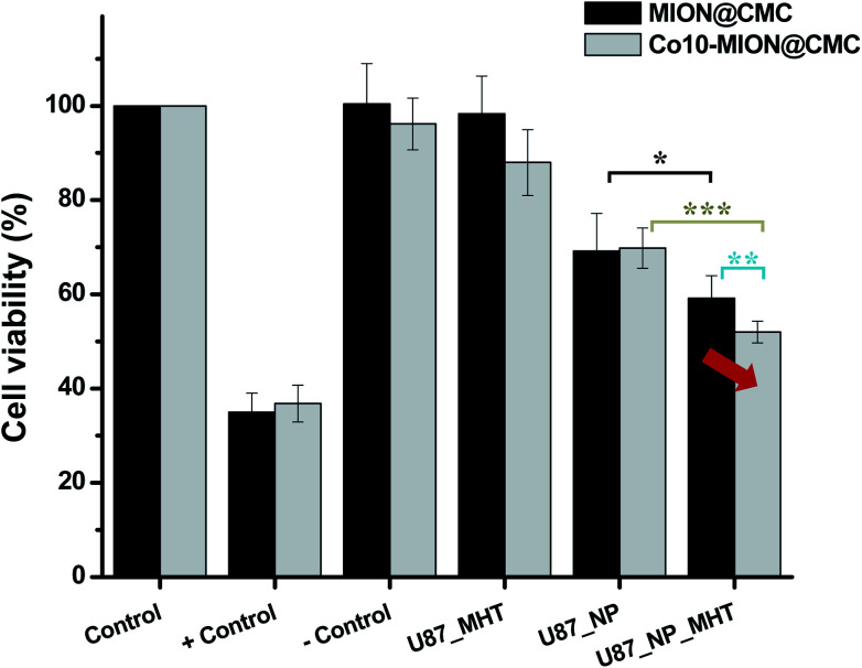
Magnetite nanoparticles are one of the most promising ferrofluids for hyperthermia applications due to the combination of unique physicochemical and magnetic properties. In this study, we designed and produced superparamagnetic ferrofluids composed of magnetite (Fe3O4, MION) and cobalt-doped magnetite (Co x -MION, x = 3, 5, and 10% mol of cobalt) nanoconjugates through an eco-friendly aqueous method using carboxymethylcellulose (CMC) as the biocompatible macromolecular ligand. The effect of the gradual increase of cobalt content in Fe3O4 nanocolloids was investigated in-depth using XRD, XRF, XPS, FTIR, DLS, zeta potential, EMR, and VSM analyses. Additionally, the cytotoxicity of these nanoconjugates and their ability to cause cancer cell death through heat induction were evaluated by MTT assays in vitro. The results demonstrated that the progressive substitution of Co in the magnetite host material significantly affected the magnetic anisotropy properties of the ferrofluids. Therefore, Co-doped ferrite (Co x Fe(3-x)O4) nanoconjugates enhanced the cell-killing activities in magnetic hyperthermia experiments under alternating magnetic field performed with human brain cancer cells (U87). On the other hand, the Co-doping process retained the pristine inverse spinel crystalline structure of MIONs, and it has not significantly altered the average nanoparticle size (ca.∼7.1 ± 1.6 nm). Thus, the incorporation of cobalt into magnetite-polymer nanostructures may constitute a smart strategy for tuning their magnetothermal capability towards cancer therapy by heat generation.


Conflict of interest statement

The authors declare no competing interest regarding the publication of this paper.

Figures

Fig. 1
Fig. 1. TEM images and histogram of the size distribution of (A) MION@CMC, (B) Co3-MION@CMC, and (C) Co5-MION@CMC.
Fig. 2
Fig. 2. (A) XRD patterns of (a) MION@CMC, (b) Co3-MION@CMC, (c) Co5-MION@CMC, and (d) Co10-MION@CMC samples compared with the reference pattern of Fe3O4 (JCPDS – 89-0691). (B) WD-XRF spectra of Co10-MION@CMC. (C) Detail of the WD-XRF spectra region associated with the range of Co peaks for (a) MION@CMC, (b) Co3-MION@CMC, (c) Co5-MION@CMC, and (d) Co10-MION@CMC.
Fig. 3
Fig. 3. XPS spectra of (A) Fe 2p and (B) Co 2p regions for (a) MION@CMC and (d) Co10-MION@CMC. (C) Spectra of Co 2p3/2 region for (a) MION@CMC, (b) Co3-MION@CMC, (c) Co5-MION@CMC, and (d) Co10-MION@CMC. Inset: evolution of intensity at Co 2p3/2 peak with increasing cobalt content.
Fig. 4
Fig. 4. XPS spectra of (A) C 1s and (B) O 1s regions obtained for (a) CMC ligand, (b) MION@CMC, and (c) Co10-MION@CMC.
Fig. 5
Fig. 5. (A) FTIR spectra of (a) MION@CMC, (b) Co3-MION@CMC, (c) Co5-MION@CMC and (d) Co10-MION@CMC. (B) Detail of the FTIR spectra region associated with the range of β1-4, Fe–O and Co–O vibrations. (C) Evolution of Fe–O and Co–O bands due to cobalt doping.
Fig. 6
Fig. 6. DLS and ZP results obtained from MION@CMC and Cox-MION@CMC at pH = 6.5 ± 0.5 and conductivity = 420 ± 10 μS (n ≥ 10; mean ± standard deviation, SD).
Fig. 7
Fig. 7. EMR spectra of (a) MION@CMC, (b) Co3-MION@CMC, (c) Co5-MION@CMC, and (d) Co10-MION@CMC for (A) samples in powder form and (B) ferrofluids. (C) EMR spectra of Co5-MION@CMC ferrofluids with (I) 0.1 mg mL−1, (II) 0.5 mg mL−1, (III) 1.0 mg mL−1 and (IV) 5.0 mg mL−1.
Fig. 8
Fig. 8. Hysteresis curves measured at (A) 301 K and (B) 77 K for (a) MION@CMC, (b) Co3-MION@CMC, (c) Co5-MION@CMC, and (d) Co10-MION@CMC. (C) Magnetization curves in the temperature range from 301 K to 77 K for zero-field cooling (ZFC) and after applying a field of 70 Oe for field cooling (FC).
Fig. 9
Fig. 9. (A) Variation in “net” temperature over time for (a) MION@CMC, (b) Co3-MION@CMC, (c) Co5-MION@CMC, and (d) Co10-MION@CMC. (B) SAR values in comparison to the coercive field of nanoconjugates at low temperature.
Fig. 10
Fig. 10. MTT in vitro assays for normal (HEK 293T, (A)) and cancer (U87, (B)) cells incubated with MION@CMC, Co3-MION@CMC, Co5-MION@CMC, and Co10-MION@CMC nanocolloids for 24 h. (C) Dose–response curves and (D) EC50 analysis (n = 6; mean ± SD).
Fig. 11
Fig. 11. (A) XRD patterns of (a) Co10-magnetite and (b) Co10-hematite powders, compared with reference patterns (Fe3O4 and α-Fe2O3). MTT assays for (B) HEK 293T and (C) U87 cells incubated with Co10-magnetite and Co10-hematite nanoparticles for 24 h. (D) Dose–response curves and (E) EC50 analysis (n = 6; mean ± SD).
Fig. 12
Fig. 12. Cell viability response of the U87 incubated with MION@CMC and Co10-MION@CMC systems (C = 15 μg mL−1) in the absence (U87-NP) and the presence of alternate magnetic field (U87-NP-MHT) (n = 6; mean ± SD; one-way ANOVA followed by Bonferroni's test with * = p < 0.05; ** = p < 0.01; *** = p < 0.001. Only the significant differences were presented, excluding comparison to/between controls).

References

    1. Myrovali E. Maniotis N. Makridis A. Terzopoulou A. Ntomprougkidis V. Simeonidis K. Sakellari D. Kalogirou O. Samaras T. Salikhov R. Spasova M. Farle M. Wiedwald U. Angelakeris M. Sci. Rep. 2016;6:1–11. doi: 10.1038/srep37934. - DOI - PMC - PubMed
    1. Calatayud M. P. Soler E. Torres T. E. Campos-Gonzalez E. Junquera C. Ibarra M. R. Goya G. F. Sci. Rep. 2017;7:1–16. doi: 10.1038/s41598-017-09059-7. - DOI - PMC - PubMed
    1. Ahmed S., Rajak B. L., Gogoi M. and Sarma H. D., in Smart Healthcare for Disease Diagnosis and Prevention, Elsevier Inc., 2020, pp. 153–173
    1. Hadadian Y. Ramos A. P. Pavan T. Z. Sci. Rep. 2019;9:1–14. doi: 10.1038/s41598-019-54250-7. - DOI - PMC - PubMed
    1. Sathya A. Guardia P. Brescia R. Silvestri N. Pugliese G. Nitti S. Manna L. Pellegrino T. Chem. Mater. 2016;28:1769–1780. doi: 10.1021/acs.chemmater.5b04780. - DOI