Review
. 2022 Sep 5:13:958442.
doi: 10.3389/fendo.2022.958442. eCollection 2022.

Dual-tracer PET/CT imaging to determine tumor heterogeneity in a patient with metastatic ACTH-secreting neuroendocrine neoplasm: A case report and literature review

Affiliations
Review

Dual-tracer PET/CT imaging to determine tumor heterogeneity in a patient with metastatic ACTH-secreting neuroendocrine neoplasm: A case report and literature review

Daria Ryzhkova et al. Front Endocrinol (Lausanne). .

Abstract

Introduction: We present a case of a patient with disseminated ACTH-secreting neuroendocrine neoplasm with biologic heterogeneity between a primary tumor and metastases. The diagnosis was obtained and multidisciplinary management was conducted with a positron emission tomography/computed tomography (PET/CT) scan with Gallium-68 [68Ga]-labeled dodecanetetraacetic acid-tyrosine-3-octreotate ([68Ga]-DOTA-TATE) and Fluor-18 [18F]-fluorodeoxyglucose ([18F]-FDG).

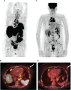
Case report: A PET/CT scan revealed a difference between [68Ga]-DOTA-TATE and [18F]-FDG uptake in primary tumor and several metastases. PET/CT showed high [18F]-FDG uptake and lack of [68Ga]-DOTA-TATE in the primary tumor, whereas both [68Ga]-DOTA-TATE and [18F]-FDG hyperaccumulation were identified in the majority of metastases. Despite positive [68Ga]-DOTA-TATE PET/CT, which is associated with high affinity with the somatostatin receptor 2 subtype, immunohistochemical examination revealed overexpression of the somatostatin receptor 5 subtype only. Perhaps, this explained the ineffectiveness of the treatment with "cold" somatostatin analogs.

Conclusion: This case had an aggressive clinical course, despite cytoreductive surgical treatment and somatostatin analog therapy. PET/CT imaging with two tracers is a molecular tool that demonstrates a biologic heterogeneity between a primary tumor and metastases and yields additional information that may influence the choice of the patient management strategy.

Keywords: PET/CT; [18F]-FDG; [68Ga]-DOTATATE; ectopic Cushing’s syndrome; ovarian NET.


Conflict of interest statement

The authors declare that the research was conducted in the absence of any commercial or financial relationships that could be construed as a potential conflict of interest. The reviewer MM declared a past collaboration with the authors DR and LM to the handling editor.

Figures

Figure 1
Figure 1
Positron emission tomography/computed tomography (PET/CT) images with Gallium-68 [68Ga]-labeled dodecanetetraacetic acid-tyrosine-3-octreotate ([68Ga]-DOTA-TATE) and Fluor-18 [18F]-fluorodeoxyglucose ([18F]-FDG). (A) The maximum intensity projection (MIP) images summarize the physiological and pathological distribution of [68Ga]-DOTA-TATE in the body. (B) PET/CT scan showed multiple mediastinal lymphatic nodes (blue dotted arrow) and widespread bone metastases with significant uptake of [68Ga]-DOTA-TATE. (C) [68Ga]-DOTA-TATE-positive bone metastasis in the left iliac wing (white arrow). (D) [68Ga]-DOTA-TATE-negative solid mass in the left ovary (red arrow). (E) The MIP images summarize the physiological and pathological distribution of [18F]-FDG in the body. (F) Multiple [68Ga]-DOTA-TATE-positive metastases in mediastinal lymphatic nodes (blue dotted arrow). (G) The axial PET/CT image demonstrated low [18F]-FDG uptake in the bone metastasis in the left iliac wing (white arrow). (H) The axial slice of [18F]-FDG PET/CT showed high [18F]-FDG uptake in the solid mass in the left ovary (red arrow).
Figure 2
Figure 2
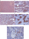
Histological and immunohistochemical examination of primary neuroendocrine tumor of the left ovary. (A) Hematoxylin and eosin staining ×50. (B) CD56 ×50. (C) ACTH ×50. (D) GLUT-1 ×100. (E) SSTR 5 subtype ×200.
Figure 3
Figure 3
PET/CT images with [68Ga]-DOTA-TATE and [18F]-FDG. (A) The maximum intensity projection (MIP) images summarize the physiological and pathological distribution of [68Ga]-DOTA-TATE in the body. PET/CT scan showed an increase in the size of the previously detected metastases and the appearance of a new mediastinal lymph node and bone metastases. (B) [68Ga]-DOTA-TATE. PET/CT revealed an avid metastatic lesion in the upper inner quadrant of the left breast (white arrow). (C) The MIP images of [18F]-FDG PET showed intense tracer uptake in the same area as the [68Ga]-DOTA-TATE PET/CT scan. (D) [18F]-FDG-positive metastatic lesion in the left breast (white arrow).
Figure 4
Figure 4
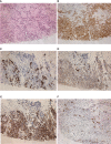
Histological and immunohistochemical examination of a metastatic lesion in the left breast. (A) Hematoxylin and eosin staining ×200. (B) Synaptophysin ×200. (C) ACTH ×200. (D) GLUT-1 ×200. (E) SSTR 5 subtype ×200. (F) Ki-67 ×200.

References

    1. Huang B, Wu X, Zhou Q, Hu Y, Zhao H, Zhu H, et al. . Cushing’s syndrome secondary to ectopic ACTH secretion from carcinoid tumor within an ovarian mature teratoma: A case report and review of the literature. Gynecol Endocrinol (2014) 30(3):192–6. doi: 10.3109/09513590.2013.871518 - DOI - PubMed
    1. Khan M, Waguespack S, Habra M, Broaddus R, Jimenez C. Acute-onset ectopic adrenocorticotropic hormone syndrome secondary to metastatic endometrioid carcinoma of the ovaries as a fatal complication. J Clin Oncol (2011) 29:e462–464. doi: 10.1200/JCO.2010.34.3848 - DOI - PubMed
    1. Sworczak K, Blaut K, Malecha M, Lewczuk A. Ectopic ACTH syndrome associated with ovarian steroid-cell tumor. J Endocrinol Invest (2002) 25:369–72. doi: 10.1007/BF03344020 - DOI - PubMed
    1. Watson J, Taylor M, Pampiglione J, Rasbridge S, Armitage M. An exception to the rule: Ectopic ACTH production from functional neuroendocrine tissue in an ovarian dermoid cyst. J Endocrinol Invest (2001) 24:602–5. doi: 10.1007/BF03343930 - DOI - PubMed
    1. Sedhom R, Hu S, Ohri A, Infantino D, Lubitz S. Symptomatic cushing’s syndrome and hyperandrogenemia in a steroid cell ovarian neoplasm: A case report. J Med Case Rep (2016) 10:278. doi: 10.1186/s13256-016-1061-x - DOI - PMC - PubMed

LinkOut - more resources