. 2022 Sep;42(5):903-914.
doi: 10.5851/kosfa.2022.e44. Epub 2022 Sep 1.

Monitoring Cellular Immune Responses after Consumption of Selected Probiotics in Immunocompromised Mice

Affiliations

Monitoring Cellular Immune Responses after Consumption of Selected Probiotics in Immunocompromised Mice

Seok-Jin Kang et al. Food Sci Anim Resour. 2022 Sep.

Abstract

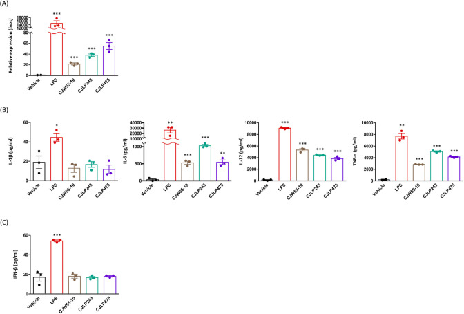
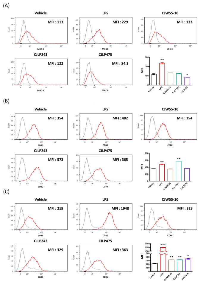
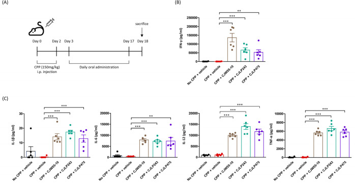
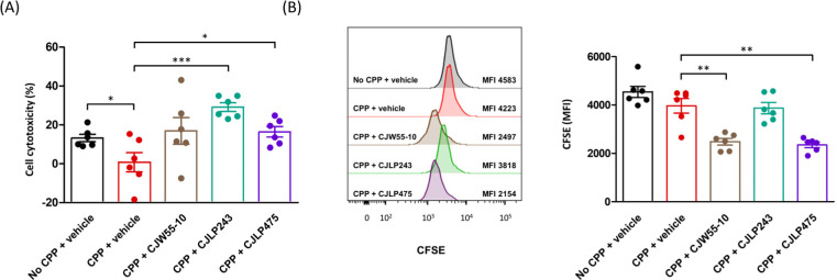
Probiotics are currently considered as one of tools to modulate immune responses under specific clinical conditions. The purpose of this study was to evaluate whether oral administration of three different probiotics (Lactiplantibacillus plantarum CJLP243, CJW55-10, and CJLP475) could evoke a cell-mediated immunity in immunodeficient mice. Before conducting in vivo experiments, we examined the in vitro potency of these probiotics for macrophage activation. After co-culture with these probiotics, bone marrow derived macrophages (BMDMs) produced significant amounts of proinflammatory cytokines including interleukin-6 (IL-6), IL-12, and tumor necrosis factor-α (TNF-α). Levels of inducible nitric oxide synthase (inos) and co-stimulatory molecules (CD80 and CD86) were also upregulated in BMDMs after treatment with some of these probiotics. To establish an immunocompromised animal model, we intraperitoneally injected mice with cyclophosphamide on day 0 and again on day 2. Starting day 3, we orally administered probiotics every day for the last 15 d. After sacrificing experimental mice on day 18, splenocytes were isolated and co-cultured with these probiotics for 3 d to measure levels of several cytokines and immune cell proliferation. Results clearly indicated that the consumption of all three probiotic strains promoted secretion of interferon-γ (IFN-γ), IL-1β, IL-6, IL-12, and TNF-α. NK cell cytotoxicity and proliferation of immune cells were also increased. Taken together, our data strongly suggest that consumption of some probiotics might induce cell-mediated immune responses in immunocompromised mice.

Keywords: animal model; immune response; probiotics.


Conflict of interest statement

Yun HS is currently employed by the CJ CheilJedang Corporation, Korea. None of the other authors had any conflict of interests.

Figures

Fig. 1.
Fig. 1.. BMDMs stimulated with selected probiotics induce inos expression and pro-inflammatory cytokine production.
BMDMs (5×10⁴ cell/well) from C57BL/6 mice were co-cultured with each heat-killed probiotics (CJW55-10, CJLP243, or CJLP475) at a density of 6×10⁶ CFU/mL. (A) Three days after co-culture, quantitative real-time RT-PCRs were performed to determine relative expression levels of inos in BMDMs. (B, C) After 3 d, quantities of pro-inflammatory cytokines (B) and IFN-β (C) in culture supernatants were determined by ELISAs. Vehicle, PBS treatment; LPS, LPS (10 μg/mL) treatment. All results are shown as means±SEM. Significant differences compared with the vehicle group are indicated as * p<0.05, ** p<0.01, *** p<0.001. BMDM, bone marrow derived macrophage; inos, inducible nitric oxide synthase; IFN, interferon; PBS, phosphate buffered saline; LPS, lipopolysaccharide.
Fig. 2.
Fig. 2.. BMDMs stimulated with selected probiotics up-regulates surface expression levels of co-stimulatory molecules such as CD80 and CD86.
BMDMs (5×10⁴ cell/well) from C57BL/6 mice were co-cultured with each heat-killed probiotics (CJW55-10, CJLP243, CJLP475) at a density of 6×10⁶ CFU/mL. Three days after co-culture, flow cytometry analyses were performed to measure surface expression levels of MHC class II (Ab) (A), CD80 (B), and CD86 (C). Gray line, before stimulation; Red line, 3 d after stimulation; Vehicle, PBS treatment; LPS, LPS (10 μg/mL) treatment. All results are shown as means±SEM. Significant differences compared with the vehicle group are indicated as * p<0.05, ** p<0.01, *** p<0.001. MFI, mean fluorescence intensity; BMDM, bone marrow derived macrophage; PBS, phosphate buffered saline; LPS, lipopolysaccharide.
Fig. 3.
Fig. 3.. Oral administration of selected probiotics induces an antigen-specific cell-mediated immunity in immunodeficient mice.
(A) Experimental scheme of CPP-induced immunocompromised mouse model. (B, C) After sacrificing mice, splenocytes (1×10⁶ cells/well) were re-stimulated with each heat-killed probiotics (CJW55-10, CJLP243, CJLP475) at density of 6×10⁶ CFU/mL for 3 d. After culture, quantities of IFN-γ (B) and pro-inflammatory cytokines (C) in culture supernatants were measured by ELISAs. No CPP+vehicle, PBS treatment without CPP administration; CPP+vehicle, PBS treatment with CPP administration; CPP+CJW55-10, CJW55-10 treatment with CPP administration; CPP+CJLP243, CJLP243 treatment with CPP administration; CPP+CJLP475, CJLP475 treatment with CPP administration. All results are shown as means±SEM. Significant differences compared with the “CPP+vehicle” group are indicated by ** p<0.01 and *** p<0.001. CPP, cyclophosphamide, IFN, interferon; PBS, phosphate buffered saline.
Fig. 4.
Fig. 4.. Oral consumption of selected probiotics increases NK cell activity and antigen-specific leukocyte proliferation in immunodeficient mice.
Experimental scheme of the CPP-induced immunocompromised mouse model is shown in Fig. 3A. (A) After sacrificing mice, splenocytes (1×10⁶ cells/well) and YAC-1 cells (1×10⁴ cell/well) (E:T ratio=100:1) were seeded to a 96-well plate in 200 μL of RPMI-10 and co-incubated at 37°C for 6 h. The cytotoxicity of target cells was then measured with an LDH assay kit. (B) After sacrificing mice, splenocytes (1×10⁶ cells/well) were labeled with CFSE (5 μM) and re-stimulated with each heat-killed probiotics (CJW55-10, CJLP243, CJLP475) at density of 6×10⁶ CFU/mL for 3 d. Relative proliferation of splenocytes was assessed by flow cytometry analyses. No CPP+vehicle, PBS treatment without CPP administration; CPP+vehicle, PBS treatment with CPP administration; CPP+CJW55-10, CJW55-10 treatment with CPP administration; CPP+CJLP243, CJLP243 treatment with CPP administration; CPP+CJLP475, CJLP475 treatment with CPP administration. All results are shown as means±SEM. Significant differences compared with the “CPP+vehicle” group are indicated by * p<0.05, ** p<0.01, and *** p<0.001. CPP, cyclophosphamide; MFI, mean fluorescence intensity; CFSE, carboxyfluorescein succinimidyl ester; PBS, phosphate buffered saline.

References

    1. Bui VT, Tseng HC, Kozlowska A, Maung PO, Kaur K, Topchyan P, Jewett A. Augmented IFN-γ and TNF-α induced by probiotic bacteria in NK cells mediate differentiation of stem-like tumors leading to inhibition of tumor growth and reduction in inflammatory cytokine release; regulation by IL-10. Front Immunol. 2015;6:576. doi: 10.3389/fimmu.2015.00576. - DOI - PMC - PubMed
    1. Bujalance C, Moreno E, Jimenez-Valera M, Ruiz-Bravo A. A probiotic strain of Lactobacillus plantarum stimulates lymphocyte responses in immunologically intact and immunocompromised mice. Int J Food Microbiol. 2007;113:28–34. doi: 10.1016/j.ijfoodmicro.2006.07.014. - DOI - PubMed
    1. Choi DW, Jung SY, Kang J, Nam YD, Lim SI, Kim KT, Shin HS. Immune-enhancing effect of nanometric Lactobacillus plantarum nF1 (nLp-nF1) in a mouse model of cyclophosphamide-induced immunosuppression. J Microbiol Biotechnol. 2018;28:218–226. doi: 10.4014/jmb.1709.09024. - DOI - PubMed
    1. Crump GM, Zhou J, Mashayekh S, Grimes CL. Revisiting peptidoglycan sensing: Interactions with host immunity and beyond. Chem Commun. 2020;56:13313–13322. doi: 10.1039/D0CC02605K. - DOI - PMC - PubMed
    1. de Melo Pereira GV, de Oliveira Coelho B, Magalhães AI, Júnior, Thomaz-Soccol V, Soccol CR. How to select a probiotic? A review and update of methods and criteria. Biotechnol Adv. 2018;36:2060–2076. doi: 10.1016/j.biotechadv.2018.09.003. - DOI - PubMed