Review
. 2022 Jul 18;4(17):3462-3478.
doi: 10.1039/d2na00420h. eCollection 2022 Aug 23.

Recent advances in mechanical force-responsive drug delivery systems

Affiliations
Review

Recent advances in mechanical force-responsive drug delivery systems

Panqin Ma et al. Nanoscale Adv. .

Abstract

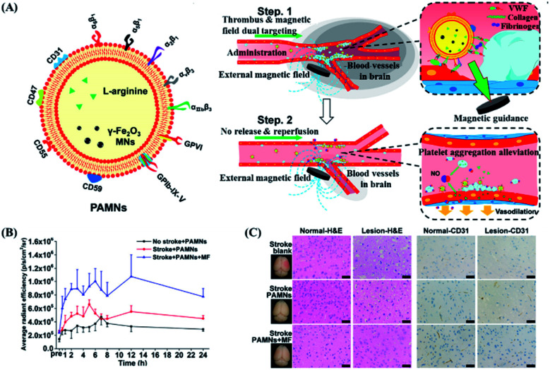
Mechanical force responsive drug delivery systems (in terms of mechanical force induced chemical bond breakage or physical structure destabilization) have been recently explored to exhibit a controllable pharmaceutical release behaviour at a molecular level. In comparison with chemical or biological stimulus triggers, mechanical force is not only an external but also an internal stimulus which is closely related to the physiological status of patients. However, although this mechanical force stimulus might be one of the most promising and feasible sources to achieve on-demand pharmaceutical release, current research in this field is still limited. Hence, this tutorial review aims to comprehensively evaluate the recent advances in mechanical force-responsive drug delivery systems based on different types of mechanical force, in terms of direct stimulation by compressive, tensile, and shear force, or indirect/remote stimulation by ultrasound and a magnetic field. Furthermore, the exciting developments and current challenges in this field will also be discussed to provide a blueprint for potential clinical translational research of mechanical force-responsive drug delivery systems.


Conflict of interest statement

There are no conflicts to declare.

Figures

Fig. 1
Fig. 1. Schematic diagram of a drug delivery system triggered by different mechanical forces. (A and B) Drug release in response to endogenous mechanical force. Reproduced with permission. Copyright 2016, American Chemical Society. (A) Drug release in response to compressive or tensile force. (i) The deformation of the carrier under compression causes the loaded drug to be released. (ii) Tension-responsive particle deformation causes the release of the encapsulated drug. (iii) The material will degrade when exposed to relevant enzymes under tension strain. (B) Drug release can be triggered by (i) aggregate dissociation or (ii) shear force caused by particle deformation. (C and D) Drug release in response to exogenous mechanical force generated by ultrasound and magnetic field. Reproduced with permission. Copyright 2020, Elsevier BV. (C) Ultrasound produces a variety of mechanical effects, including push/pull, acoustic radiation force, and microjets, which can cause the short opening of blood vessels and control the release of drugs. (D) The magnetic field can destroy the endothelial cell–cell connection to achieve controlled drug release. (E) Drug release in response to cellular forces. Reproduced with permission. Copyright 2020, Royal Society of Chemistry.
Scheme 1
Scheme 1. Schematic illustration of a mechanical force responsive DDS, upon the stimulation of endogenous mechanical force (compression, tension, and shear force) or exogenous mechanical force (ultrasound and magnetism).
Fig. 2
Fig. 2. Mechanical activity and stability of a mechanically activatable microcapsule (MAMC). (A) The volume (confocal, scale bar = 50 μm) and shape (SEM, scale bar = 50 μm) of fast-degrading MAMCs containing dextran (green) with labeled shells (red) and illustration of the rupture mechanism. (B) The stability and the mechanical activation response at low pressure of slow degrading MAMCs from day 1 (D1) to incubation for more than 70 days (D70). Fluorescent images scale bar = 100 μm. (C) Typical confocal images of MAMCs in different stiffness matrix hydrogels (matrix: blue, MAMCs: green, ruptured MAMCs with loss of internal signal). Scale bar = 200 μm. Reproduced with permission. Copyright 2019, John Wiley & Sons.
Fig. 3
Fig. 3. Correlation between the amount of drug released and exposed region of the stretched film. (A)Scanning electron microscope (SEM) images and energy dispersive X-ray spectroscopy images of the stretched membranes at various tensile strains. (B) Profiles of the relative released drug versus strain and the ‘ratio of the exposed region’ under experimental and theoretical conditions. Reproduced with permission. Copyright 2018, Elsevier BV.
Fig. 4
Fig. 4. Structure and drug release of shear-reactive polymer vesicles. (A) Schematic diagram of the inner cavity of polymer vesicles with or without shear stress. (B) Fluorescence microscope images of polymer vesicles filled with sodium fluorescein with or without shear stress obtained using a microfluidic device with multiple contractions from left to right. (C) Fluorescein sodium is released from the shear-responsive polymer vesicles with a shear flow (black) or in the absence of a shear flow (red), and non-shear-responsive polymer vesicles with a shear flow (blue). Reproduced with permission. Copyright 2021, John Wiley & Sons.
Fig. 5
Fig. 5. Schematic illustration of the interaction between functionalized polystryrene nanoparticles (as a vector targeting endothelial cells or VCT) and endothelial cells under high shear stress. Schematic diagram of (A) leukocytes and (B) VTC that mimics white blood cells activating endothelial cells under the blood flow mediated by overexpression of ICAM 1 and E-selectin. (C) In microfluidics, the channel controls the flow of injection pump with ability to apply shear stress on endothelial cells. Reproduced with permission. Copyright 2020, Wiley VCH.
Fig. 6
Fig. 6. In vitro drug release experiment of US-responsive DOX–(TA/PVPON-58)8 capsules. (A) On-off controllable drug release of ultrasound-responsive capsules of DOX–(TA/PVPON-58)8 capsules. Arrows indicate the point at which the US was applied (ON). (B) The dependence of capsule rupture (%) on the US power intensity (W cm−2) under the condition of 20 seconds of US duration time, pH = 7.4. (C) In vitro anticancer activity of ultrasound-responsive DOX–(TA/PVPON-58)8 capsules. (D) The dependence of capsule rupture (%) on the time of ultrasonic treatment under the condition of a US intensity of 14 W cm−1, pH = 7.4. Reproduced with permission. Copyright 2017, American Chemical Society.
Fig. 7
Fig. 7. Polymer perfluoropentane nanoemulsion as an ultrasound-responsive delivery system for encapsulating hydrophobic drugs. (A) Schematic of polymer perfluoropentane nanoemulsion production and ultrasonic drug uncaging. (B) Polydispersity index of perfluoropentane nanoemulsion loaded with propofol (PPF), nicardipine (NIC), verapamil (VER), dexmedetomidine (DEX), ketamine (KET), doxorubicin (DOX) or cisplatin. (C) Stability of perfluoropentane nanoemulsion under frozen storage and thawing. (D) In vitro drug release under ultrasound stimulation of perfluoropentane nanoemulsion loaded with different hydrophobic drugs (650 kHz ultrasound treatment, 50 ms pulse repetition 60 times). Reproduced with permission. Copyright 2019, Elsevier Ltd.
Fig. 8
Fig. 8. Magnetic response PAMN for in vivo verification of early ischemic stroke. (A) Schematic diagram of the structure of PAMN and the effect of in vivo magnetic targeted drug release. (B) Quantitative analysis of the average DiR fluorescence signal intensity shows that the magnetic field can increase the distribution of the drug in the lesion area. (C) H&E staining and CD31 staining of normal brain (left) tissue and ischemic lesion (right) tissue under different treatment conditions (scale bar: 20 μm). Reproduced with permission. Copyright 2020, American Chemical Society.
Fig. 9
Fig. 9. Implantable, battery-free magnetically responsive pump as a validation of a drug delivery system. (A) In vitro performance test of drug delivery stability of the magnetic response pump. (B) Testing the biocompatibility of the magnetic response pump in different parts by H&E and MT staining. The scale bars are 100 μm. (C) In vivo pharmacodynamic experiment of insulin changes caused by in vivo administration of exenatide-loaded magnetic response pump infusion per actuation with up to 300 consecutive actuations. Reproduced with permission. Copyright 2020, Elsevier.
None
Panqin Ma
None
Zibiao Li
None
Yun-Long Wu

Similar articles

Cited by

References

    1. Zhang Y. Yu J. Bomba H. N. Zhu Y. Gu Z. Mechanical Force-Triggered Drug Delivery. Chem. Rev. 2016;116(19):12536–12563. doi: 10.1021/acs.chemrev.6b00369. - DOI - PubMed
    1. Rozenbaum R. T. Andrén O. C. J. van der Mei H. C. Woudstra W. Busscher H. J. Malkoch M. Sharma P. K. Penetration and Accumulation of Dendrons with Different Peripheral Composition in Pseudomonas aeruginosa Biofilms. Nano Lett. 2019;19(7):4327–4333. doi: 10.1021/acs.nanolett.9b00838. - DOI - PMC - PubMed
    1. Albuquerque L. J. C. Sincari V. Jäger A. Kucka J. Humajova J. Pankrac J. Paral P. Heizer T. Janouškova O. Davidovich I. Talmon Y. Pouckova P. Štěpánek P. Sefc L. Hruby M. Giacomelli F. C. Jäger E. pH-responsive polymersome-mediated delivery of doxorubicin into tumor sites enhances the therapeutic efficacy and reduces cardiotoxic effects. J. Controlled Release. 2021;332:529–538. doi: 10.1016/j.jconrel.2021.03.013. - DOI - PubMed
    1. Zhang W. Wen Y. He D.-X. Wang Y.-F. Liu X.-L. Li C. Liang X.-J. Near-infrared AIEgens as transformers to enhance tumor treatment efficacy with controllable self-assembled redox-responsive carrier-free nanodrug. Biomaterials. 2019;193:12–21. doi: 10.1016/j.biomaterials.2018.12.007. - DOI - PubMed
    1. Yang J. Pan S. Gao S. Dai Y. Xu H. Anti-recurrence/metastasis and chemosensitization therapy with thioredoxin reductase-interfering drug delivery system. Biomaterials. 2020;249:120054. doi: 10.1016/j.biomaterials.2020.120054. - DOI - PubMed