. 2022 Aug 24;9(9):412.
doi: 10.3390/bioengineering9090412.

Optimized Decellularization Protocol for Large Peripheral Nerve Segments: Towards Personalized Nerve Bioengineering

Affiliations

Optimized Decellularization Protocol for Large Peripheral Nerve Segments: Towards Personalized Nerve Bioengineering

Alois Hopf et al. Bioengineering (Basel). .

Abstract

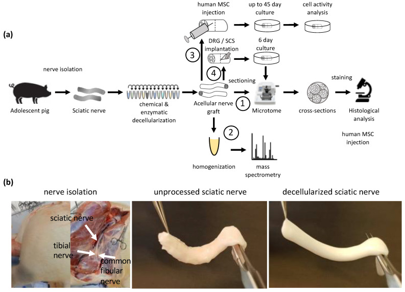
Nerve injuries remain clinically challenging, and allografts showed great promise. Decellularized nerve allografts possess excellent biocompatibility and biological activity. However, the vast majority of decellularization protocols were established for small-size rodent nerves and are not suitable for clinical application. We aimed at developing a new method of decellularizing large-diameter nerves suitable for human transplantation. Repeated rounds of optimization to remove immunogenic material and preserve the extracellular structure were applied to the porcine sciatic nerve. Following optimization, extensive in vitro analysis of the acellular grafts via immunocytochemistry, immunohistology, proteomics and cell transplantation studies were performed. Large segments (up to 8 cm) of the porcine sciatic nerve were efficiently decellularized and histology, microscopy and proteomics analysis showed sufficient preservation of the extracellular matrix, with simultaneous consistent removal of immunogenic material such as myelin, DNA and axons, and axonal growth inhibitory molecules. Cell studies also demonstrated the suitability of these acellular grafts for 3D cell culture studies and translation to future large animal studies and clinical trials. By using non-human donors for peripheral nerve transplantation, significant drawbacks associated with the gold standard can be eliminated while simultaneously preserving the beneficial features of the extracellular matrix.

Keywords: acellular allograft; decellularization; large-gap repair; neurotmesis; peripheral nerve injuries; porcine sciatic nerve.


Conflict of interest statement

The authors declare no conflict of interest.

Figures

Figure 1
Figure 1
Experimental overview: (a) Illustration of the experimental setup. Sciatic and common fibular nerves of adolescent pigs were isolated. Following isolation, the nerves were treated with a series of various chemical and enzymatic detergents for decellularization. The resulting acellular nerve grafts were analyzed and tested in different ways. (1) Cross-sections via microtome were made from acellular graft and used for immunohistochemistry. (2) Proteomic analysis of the acellular graft to detect the number of removed peptides and known inhibitors of axonal regeneration due to the decellularization process. (3) Acellular grafts were used as 3D scaffolds for cell culture. Cells in the 3D scaffold were cultured for up to 45 days and cellular behavior was analyzed by measuring cellular metabolism via resazurin. (4) Dorsal root ganglion (DRG) and Spinal cord segments (SCS) were implanted in acellular grafts and cultured for 6 days prior to fixation, sectioning and histological analysis; (b) Representative images of nerve isolation from adolescent pigs. Unprocessed sciatic nerve with excessive fat and connective tissue and a reddish color, Decellularized sciatic nerve with removed excessive tissue and white appearance.
Figure 2
Figure 2
Removal of immunogenic material and ECM preservation by optimized protocols from large diameter pig nerves. First row: scale bar 200 μm, representative images of remaining axonal β-tubulin III, MBP and DAPI. Second row: scale bar 100 μm, zoomed-in representative images of remaining axonal β-tubulin III, MBP and DAPI. Third row: scale bar 200 μm, representative images of remaining ECM laminin structure and DAPI. Fourth row: scale bar 100 μm, zoomed-in representative images of remaining ECM laminin structure and DAPI. Each column shows a further optimization step towards protocol 5, an acellular graft with removed axonal β-tubulin III, MBP, DAPI and a conserved laminin structure signal. Protocol 5 was used for further analysis.
Figure 3
Figure 3
Quantification of remaining cellular components in longer nerve segments. (a) The remaining immunogenic material and laminin structure were analyzed every 1 cm up to the middle of an 8 cm-long pig sciatic nerve segment decellularized by protocol 5. Representative images: the remaining ECM laminin structure and DAPI are shown in the upper row, together with the remaining axonal β-tubulin III, MBP and DAPI signals in the bottom row (scale: 200 μm). (b) Graphic documentation: the remaining axonal β-tubulin III, MBP, DAPI and laminin signals were quantified. Fascicles were manually segmented and the remaining signals were normalized to signal unprocessed tissue. For statistical analysis, a one-way ANOVA was performed. Adjusted p-value: < 0.0001 = ****. ns: no significant differences. (c) Numerical documentation: percentage of the remaining axonal β-tubulin III, MBP, DAPI and laminin signals for 4 cm and 8 cm-long segments every 1 cm until the middle.
Figure 4
Figure 4
Histological analysis of the ECM and the remaining myelin structure of protocol 5. Luxol fast blue and Cresyl violet stain: myelin (blue) and nuclei (purple) (top). Hematoxylin and eosin stain: ECM (pink) and nuclei (purple) (bottom). The 5 μm thick cross-sections and longitudinal sections zoomed into framed segments. Scale: 1 mm, 200 μm, 50 μm.
Figure 5
Figure 5
Peptide removal in acellular grafts. Each column represents the protein abundance of one replicate. Ctrl 1–4 are unprocessed samples. DC 1–4 are decellularized samples. (a) Heatmap. In total, 4054 proteins were measured. Each line represents one UniProt ID identifying one protein. green: high abundance of protein in the sample. White: low abundancy of protein in the sample. (b) Total number of proteins above detection limit. (c) Number of peptides per subcategory. Subcategories defined by AmiGO2 GO class for sus scrofa and UniPort as contributor. The bar plot shows the number of detectable peptides in all replicates in chosen subcategories (cytoskeleton, axon-, axon regeneration, regulation of neuron projection development-, ECM-, collagen-containing ECM, myelin sheath associated peptides). For statistical analysis, Two-way ANOVA was performed. Adjusted p-value: <0.0001 = ****, p < 0.001 = ***, p < 0.01 = **, p < 0.05 = * ns: no significant differences.
Figure 6
Figure 6
Decellularized nerve as 3D-scaffold for hASC culture. (a) 250k hASC were injected per 3D acellular graft (dots), 10k hASC seeded in 2D-culture well as a control (triangles). Cellular activity was analyzed using resazurin every 3–5 days in media supplemented by 10% FCS (green) or 5% HPL and 2 U/mL heparin (orange). Cell activity normalized to cell activity at day 1 post-transplantation. Triangles represent cell activity in 2D-culture plates and dots represent cell activity in 3D-culture. (b) cell activity regarding FCS- and HPL-supplemented media. Relative cellular activity of hASC cultured in HPL-supplemented media normalized to relative cellular activity of hASC cultured in FCS-supplemented media. Black triangles represent normalized cell activity in 2D culture and blue dots represent normalized cell activity in 3D.

References

    1. Daly W.T., Yao L., Abu-rub M.T., O’Connell C., Zeugolis D.I., Windebank A.J., Pandit A.S. The Effect of Intraluminal Contact Mediated Guidance Signals on Axonal Mismatch during Peripheral Nerve Repair. Biomaterials. 2012;33:6660–6671. doi: 10.1016/j.biomaterials.2012.06.002. - DOI - PubMed
    1. Navarro X., Vivó M., Valero-Cabré A. Neural Plasticity after Peripheral Nerve Injury and Regeneration. Prog. Neurobiol. 2007;82:163–201. doi: 10.1016/j.pneurobio.2007.06.005. - DOI - PubMed
    1. Griffin J.W., Hogan M.V., Chhabra A.B., Deal D.N. Peripheral Nerve Repair and Reconstruction. J. Bone Jt. Surg. 2013;95:2144–2151. doi: 10.2106/JBJS.L.00704. - DOI - PubMed
    1. Sunderland S. Factors Influencing the Course of Regeneration and the Quality of the Recovery after Nerve Suture. Brain. 1952;75:19–54. doi: 10.1093/brain/75.1.19. - DOI - PubMed
    1. Pfister B.J., Gordon T., Loverde J.R., Kochar A.S., Mackinnon S.E., Cullen D.K. Biomedical Engineering Strategies for Peripheral Nerve Repair: Surgical Applications, State of the Art, and Future Challenges. Crit. Rev. Biomed. Eng. 2011;39:81–124. doi: 10.1615/CritRevBiomedEng.v39.i2.20. - DOI - PubMed

Grants and funding

LinkOut - more resources