. 2022 Sep 14;9(9):469.
doi: 10.3390/bioengineering9090469.

Virtual Scoliosis Surgery Using a 3D-Printed Model Based on Biplanar Radiographs

Affiliations

Virtual Scoliosis Surgery Using a 3D-Printed Model Based on Biplanar Radiographs

Aurélien Courvoisier et al. Bioengineering (Basel). .

Abstract

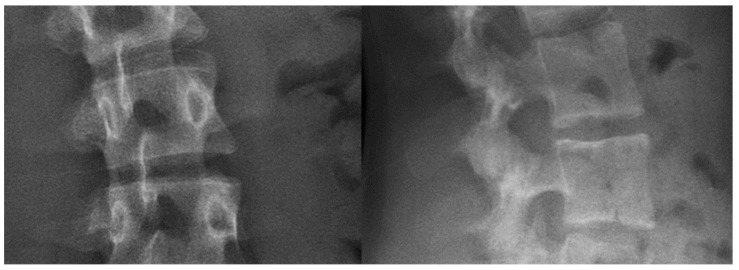
The aim of this paper is to describe a protocol that simulates the spinal surgery undergone by adolescents with idiopathic scoliosis (AIS) by using a 3D-printed spine model. Patients with AIS underwent pre- and postoperative bi-planar low-dose X-rays from which a numerical 3D model of their spine was generated. The preoperative numerical spine model was subsequently 3D printed to virtually reproduce the spine surgery. Special consideration was given to the printing materials for the 3D-printed elements in order to reflect the radiopaque and mechanical properties of typical bones most accurately. Two patients with AIS were recruited and operated. During the virtual surgery, both pre- and postoperative images of the 3D-printed spine model were acquired. The proposed 3D-printing workflow used to create a realistic 3D-printed spine suitable for virtual surgery appears to be feasible and reliable. This method could be used for virtual-reality scoliosis surgery training incorporating 3D-printed models, and to test surgical instruments and implants.

Keywords: 3D printing; additive manufacturing; bi-planar X-rays; scoliosis; virtual surgery.


Conflict of interest statement

Authors J.S., P.D., C.A., Antonio Cebrian, B.A. are full time employees of the company EOS Imaging, an affiliated company of Alphatec Holdings, Inc. Author LT is a full-time employee of the company eCential Robotics.

Figures

Figure 1
Figure 1
Process for each patient.
Figure 2
Figure 2
Grey levels of 1-mm-thick sample plates with different materials: polyactic acid with 50% ceramic (left), polyactic acid filled with 80% copper (middle), and polyactic acid (right).
Figure 3
Figure 3
Bi-planar X-rays of three 3D-printed vertebrae, each resulting from different printer settings. From top to bottom: quarter cubic 45°, hole’s infill, and personalized vertical grid 5°.
Figure 4
Figure 4
Bi-planar X-rays of the spine of an actual patient.
Figure 5
Figure 5
3D-printed shell (red) linking the vertebrae and positioned for surgery on a specifically designed foam.
Figure 6
Figure 6
3D-printed simplified pelvis and support structure.
Figure 7
Figure 7
Picture and oblique and superior X-rays of a 3D-printed vertebra with two pedicle screws inserted.
Figure 8
Figure 8
Bi-planar X-rays of patient 1 and 3D-printed twin 1. (A): Preoperative X-rays of patient 1 ((A1): frontal view, (A2): lateral view). (B): Postoperative X-rays of patient 1 ((B1): frontal view, (B2): lateral view). (C): Preoperative X-rays of 3D-printed twin 1 ((C1): frontal view, (C2): lateral view). (D): Postoperative X-rays of 3D-printed twin 1 ((D1): frontal view, (D2): lateral view).
Figure 9
Figure 9
Pictures of posterior view of 3D-printed twin 2. On the left, preoperative 3D-printed twin in prone position without pelvis and support structure. On the right, 3D-printed twin after surgery in a standing position.
Figure 10
Figure 10
Bi-planar X-rays of patient 2 and 3D-printed twin 2. (A): Preoperative X-rays of patient 2 ((A1): frontal view, (A2): lateral view). (B): Postoperative X-rays of patient 2 ((B1): frontal view, (B2): lateral view). (C): Preoperative X-rays of 3D-printed twin 2 ((C1): frontal view, (C2): lateral view). (D): Postoperative X-rays of 3D-printed twin 2 ((D1): frontal view, (D2): lateral view).

References

    1. Habib A., Jovanovich N., Muthiah N., Alattar A., Alan N., Agarwal N., Ozpinar A., Hamilton D.K. 3D printing applications in spine surgery: An evidence-based assessment toward personalized patient care. Eur. Spine J. 2022;31:1682–1690. doi: 10.1007/s00586-022-07250-7. - DOI - PubMed
    1. Wu A.M., Lin J.L., Kwan K.Y.H., Wang X.Y., Zhao J. 3D-printing techniques in spine surgery: The future prospects and current challenges. Expert Rev. Med. Devices. 2018;15:399–401. doi: 10.1080/17434440.2018.1483234. - DOI - PubMed
    1. Wong R.M.Y., Wong P.Y., Liu C., Chung Y.L., Wong K.C., Tso C.Y., Chow S.K., Cheung W.H., Yung P.S., Chui C.S., et al. 3D printing in orthopaedic surgery: A scoping review of randomized controlled trials. Bone Jt. Res. 2021;10:807–819. doi: 10.1302/2046-3758.1012.BJR-2021-0288.R2. - DOI - PMC - PubMed
    1. Yu Y., Kang J., Kim N., Heo S. Accuracy of a patient-specific 3D-printed drill guide for placement of bicortical screws in atlantoaxial ventral stabilization in dogs. PLoS ONE. 2022;17:e0272336. doi: 10.1371/journal.pone.0272336. - DOI - PMC - PubMed
    1. Xin X., Wang F., Liu X. A 3D-printed Personalized, Combined, Modular Pedicle Subtraction Osteotomy Guide Plate System: An Experimental Study. Spine. 2022;47:931–937. doi: 10.1097/BRS.0000000000004229. - DOI - PubMed

LinkOut - more resources