Review
. 2022 Sep 1;10(3):37.
doi: 10.3390/jdb10030037.

Sculpting an Embryo: The Interplay between Mechanical Force and Cell Division

Affiliations
Review

Sculpting an Embryo: The Interplay between Mechanical Force and Cell Division

Nawseen Tarannum et al. J Dev Biol. .

Abstract

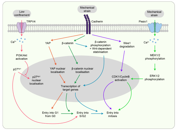
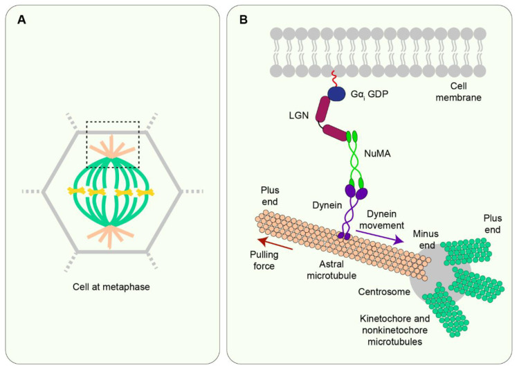
The journey from a single fertilised cell to a multicellular organism is, at the most fundamental level, orchestrated by mitotic cell divisions. Both the rate and the orientation of cell divisions are important in ensuring the proper development of an embryo. Simultaneous with cell proliferation, embryonic cells constantly experience a wide range of mechanical forces from their surrounding tissue environment. Cells must be able to read and respond correctly to these forces since they are known to affect a multitude of biological functions, including cell divisions. The interplay between the mechanical environment and cell divisions is particularly crucial during embryogenesis when tissues undergo dynamic changes in their shape, architecture, and overall organisation to generate functional tissues and organs. Here we review our current understanding of the cellular mechanisms by which mechanical force regulates cell division and place this knowledge within the context of embryogenesis and tissue morphogenesis.

Keywords: biomechanics; cell division; cell division orientation; cell division rate; cell shape; embryogenesis; mechanical force; mitosis; mitotic spindle; morphogenesis.


Conflict of interest statement

The authors declare no conflict of interest.

Figures

Figure 1
Figure 1
Mechanical force transduction at adherens junctions. Forces are transmitted through adherens junctions by cadherin dimers. The cytosolic tail of cadherin binds to p120 and β-catenin while the extracellular domain crosslinks with cadherin from the neighbouring cell. (A) In the absence of an external force, β-catenin is bound by α-catenin in its “closed” conformation to form the α-catenin-β-catenin-cadherin complex. In the “closed” state the vinculin-binding site on α-catenin is not accessible. (B) When the cell experiences a force, α-catenin undergoes a conformation change into the “open” state, thereby unmasking the vinculin-binding site and leading to vinculin recruitment at the junctions [10,11]. (C) Following vinculin recruitment, intracellular forces are generated by actomyosin contractility due to the movement of myosin head domains on F-actin filaments [12,13]. The cell that generates contractile forces acts as a force donor, and force is transmitted to the receiver cell via the junctions, thereby exerting pulling forces on the receiver cell. In addition to acting as a downstream responder to external forces, actomyosin contractility itself generates forces which act on cadherin junctions and elicit cadherin mechanotransduction in the receiver cell.
Figure 2
Figure 2
Building forces from cells to tissues. Forces generated within a cell can be transmitted to its interconnected neighbours. Cells can be modelled as tensegrity structures and tissues are, therefore, a collection of tensegrity structures which are inherently mechanosensitive [14,15,16].
Figure 3
Figure 3
The effect of cell divisions on tissue organisation. (A) In symmetric divisions, spindle alignment parallel to the plane of the tissue generates identical, side-by-side daughter cells. (B) In asymmetric divisions, the spindle aligns perpendicular to the tissue plane and divisions generate daughter cells with different cell fates, which aid tissue stratification. (C) Symmetric divisions that orient uniformly within the tissue plane lead to tissue growth in all directions. However, if the spindles align in the same direction, tissue elongation occurs along the global axis of divisions.
Figure 4
Figure 4
Contribution of cell shape and force in spindle orientation. (A) Cells placed in a microfabricated chamber assume the shape of the chamber but do not always follow the long axis rule of division. (B) Attachment of cells to the ECM via retraction fibres conserves the placement of cortical cues enabling division with the long axis of shape in rounded mitotic cells. (C) Using laser ablation to cut some retraction fibres leads to spindle alignment with cortical cues defined by the remaining retraction fibres.
Figure 5
Figure 5
Transmembrane proteins sense mechanical forces to influence cell proliferation. Mechanosensitive channels such as Piezo1 and TRPV4 as well as cadherins act as signalling centres to influence cell divisions via control of the cell cycle at different checkpoints. Coloured arrows indicate different pathways.
Figure 6
Figure 6
The spindle orientation machinery in vertebrates. (A) A schematic of a cell at metaphase is shown. The region selected by the black box is enlarged in B. (B) At interphase, NuMA is restricted within the nucleus but at the onset of mitosis, NuMA is carried to the minus end of microtubules (near the centrosomes) by dynein. Near this region, NuMA binds to LGN which, in turn, binds to Gαi GDP which is tethered to the membrane. This forms the cortically localised NuMA/dynein-LGN- Gαi complex. Dynein, due to its minus end-directed mobility, walks along astral microtubules towards the centrosomes. However, cortical localisation of the protein complex opposes dynein motion thereby exerting pulling forces on astral microtubules. These pulling forces then help to orient the mitotic spindle [74].
Figure 7
Figure 7
Mechanical forces direct the localisation of key cortical proteins to influence spindle orientation. Upon experiencing mechanical strain, cadherins promote the localisation of LGN to cell-cell contacts to regulate spindle orientation. Another spindle orientation core protein, NuMA, also localises to the cortex and potentially interacts with actin-binding proteins to influence mechanosensitive spindle positioning.

References

    1. Campinho P., Behrndt M., Ranft J., Risler T., Minc N., Heisenberg C.P. Tension-oriented cell divisions limit anisotropic tissue tension in epithelial spreading during zebrafish epiboly. Nat. Cell Biol. 2013;15:1405–1414. doi: 10.1038/ncb2869. - DOI - PubMed
    1. Fletcher G.C., Diaz-de-la-Loza M.D., Borreguero-Muñoz N., Holder M., Aguilar-Aragon M., Thompson B.J. Mechanical strain regulates the Hippo pathway in Drosophila. Development. 2018;145:dev159467. doi: 10.1242/dev.159467. - DOI - PMC - PubMed
    1. Nerurkar N.L., Mahadevan L., Tabin C.J. BMP signaling controls buckling forces to modulate looping morphogenesis of the gut. Proc. Natl. Acad. Sci. USA. 2017;114:2277–2282. doi: 10.1073/pnas.1700307114. - DOI - PMC - PubMed
    1. Scarpa E., Finet C., Blanchard G.B., Sanson B. Actomyosin-Driven Tension at Compartmental Boundaries Orients Cell Division Independently of Cell Geometry In Vivo. Dev. Cell. 2018;47:727–740.e726. doi: 10.1016/j.devcel.2018.10.029. - DOI - PMC - PubMed
    1. Tang Z., Hu Y., Wang Z., Jiang K., Zhan C., Marshall W.F., Tang N. Mechanical Forces Program the Orientation of Cell Division during Airway Tube Morphogenesis. Dev. Cell. 2018;44:313–325.e315. doi: 10.1016/j.devcel.2017.12.013. - DOI - PubMed

LinkOut - more resources