Review
. 2022 Sep 1;8(9):236.
doi: 10.3390/jimaging8090236.

Dual-Energy CT of the Heart: A Review

Affiliations
Review

Dual-Energy CT of the Heart: A Review

Serena Dell'Aversana et al. J Imaging. .

Abstract

Dual-energy computed tomography (DECT) represents an emerging imaging technique which consists of the acquisition of two separate datasets utilizing two different X-ray spectra energies. Several cardiac DECT applications have been assessed, such as virtual monoenergetic images, virtual non-contrast reconstructions, and iodine myocardial perfusion maps, which are demonstrated to improve diagnostic accuracy and image quality while reducing both radiation and contrast media administration. This review will summarize the technical basis of DECT and review the principal cardiac applications currently adopted in clinical practice, exploring possible future applications.

Keywords: applications; cardiac; dual-energy CT; review.


Conflict of interest statement

The authors declare no conflict of interest.

Figures

Figure 1
Figure 1
Illustrations showing the main DECT scanning techniques in clinical use. (A) Dual-source DECT, consisting of two-source X-ray tubes and the corresponding detectors. (B) Single-source DECT consisting of a single tube with rapid kVp switching. (C) Detector-based DECT consisting of a single source and a dual-layer detector to obtain low- and high-energy spectra.
Figure 2
Figure 2
Late iodine enhancement dual-source DECT images in a 72-year-old man. Sub-endocardial hyperenhancement (red arrows) can be seen in the left ventricular septal wall and apex at different virtual monochromatic image values (AD).
Figure 3
Figure 3
Retrospective application of VMI (AD) by dual-source DECT in a patient where the contrast bolus was mistimed in amount; low keV reconstruction increases the attenuation of iodinated contrast material, allowing for greater contrast-to-noise ratio.
Figure 4
Figure 4
Calcium scoring from coronary dual-source DECT angiography. Virtual non-contrast (A) and the corresponding DECT (B) images in a 56-year-old man with a virtual Agatston score of 73 on LAD.
Figure 5
Figure 5
Coronary dual-source DECT: (A) Left circumflex coronary artery shows severe proximal stenosis due to the presence of a calcified plaque. (B) Iodine perfusion map depicts the perfusion defect of the inferolateral wall (arrow) corresponding to a myocardial scar (arrow) in the short axis LGE MR sequence (C), suggestive of necrosis.
Figure 6
Figure 6
Coronary dual-source DECT in a 65-year-old man presenting with chest pain. (A) Automatically generated curved multiplanar reformation of the right coronary artery demonstrates greater than 75% stenosis (green asterisk). (B) Plaque analysis using monochromatic coronary reconstruction showing a clear separation of quantitative values (HU) at low keV (red asterisk: calcific plaque; green asterisk: fibrous plaque).

References

    1. Siegel M.J., Kaza R.K., Bolus D.N., Boll D.T., Rofsky N.M., De Cecco C.N., Foley W.D., Morgan D.E., Schoepf U.J., Sahani D.V., et al. White Paper of the Society of Computed Body Tomography and Magnetic Resonance on Dual-Energy CT, Part 1: Technology and Terminology. J. Comput. Assist. Tomogr. 2016;40:841–845. doi: 10.1097/RCT.0000000000000531. - DOI - PubMed
    1. De Cecco C.N., Darnell A., Rengo M., Muscogiuri G., Bellini D., Ayuso C., Laghi A. Dual-energy CT: Oncologic applications. Am. J. Roentgenol. 2012;199:S98–S105. doi: 10.2214/AJR.12.9207. - DOI - PubMed
    1. Vliegenthart R., Pelgrim G.J., Ebersberger U., Rowe G.W., Oudkerk M., Schoepf U.J. Dual-energy CT of the heart. Am. J. Roentgenol. 2012;199:S54–S63. doi: 10.2214/AJR.12.9208. - DOI - PubMed
    1. Albrecht M.H., Bickford M.W., Nance J.W.J., Zhang L., De Cecco C.N., Wichmann J.L., Vogl T.J., Schoepf U.J. State-of-the-Art Pulmonary CT Angiography for Acute Pulmonary Embolism. Am. J. Roentgenol. 2017;208:495–504. doi: 10.2214/AJR.16.17202. - DOI - PubMed
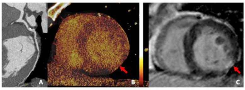
    1. Ruzsics B., Lee H., Powers E.R., Flohr T.G., Costello P., Schoepf U.J. Images in cardiovascular medicine. Myocardial ischemia diagnosed by dual-energy computed tomography: Correlation with single-photon emission computed tomography. Circulation. 2008;117:1244–1245. doi: 10.1161/CIRCULATIONAHA.107.745711. - DOI - PubMed

LinkOut - more resources