. 2022 Aug 24;8(9):899.
doi: 10.3390/jof8090899.

Evidencing New Roles for the Glycosyl-Transferase Cps1 in the Phytopathogenic Fungus Botrytis cinerea

Affiliations

Evidencing New Roles for the Glycosyl-Transferase Cps1 in the Phytopathogenic Fungus Botrytis cinerea

Matthieu Blandenet et al. J Fungi (Basel). .

Abstract

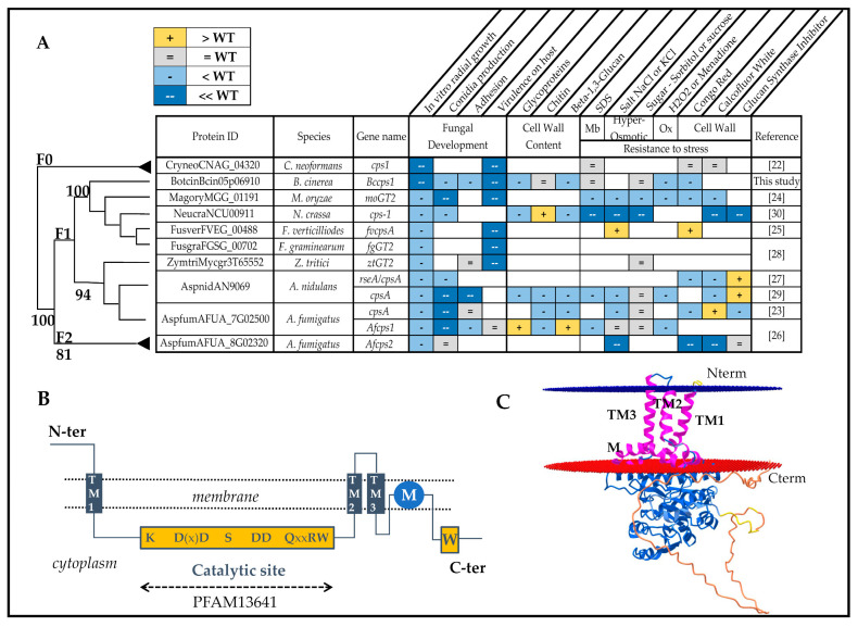
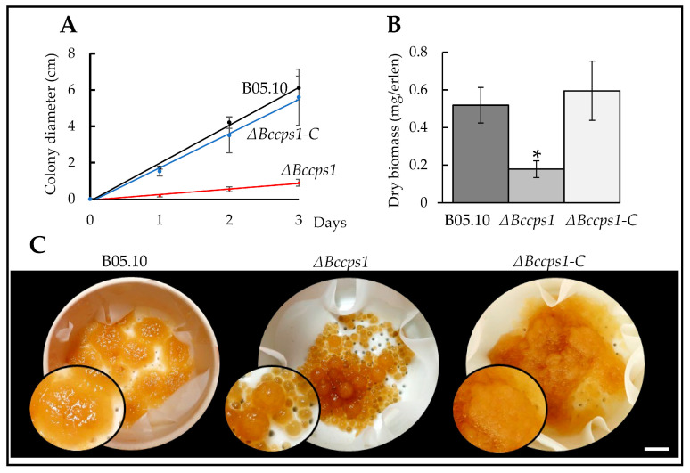
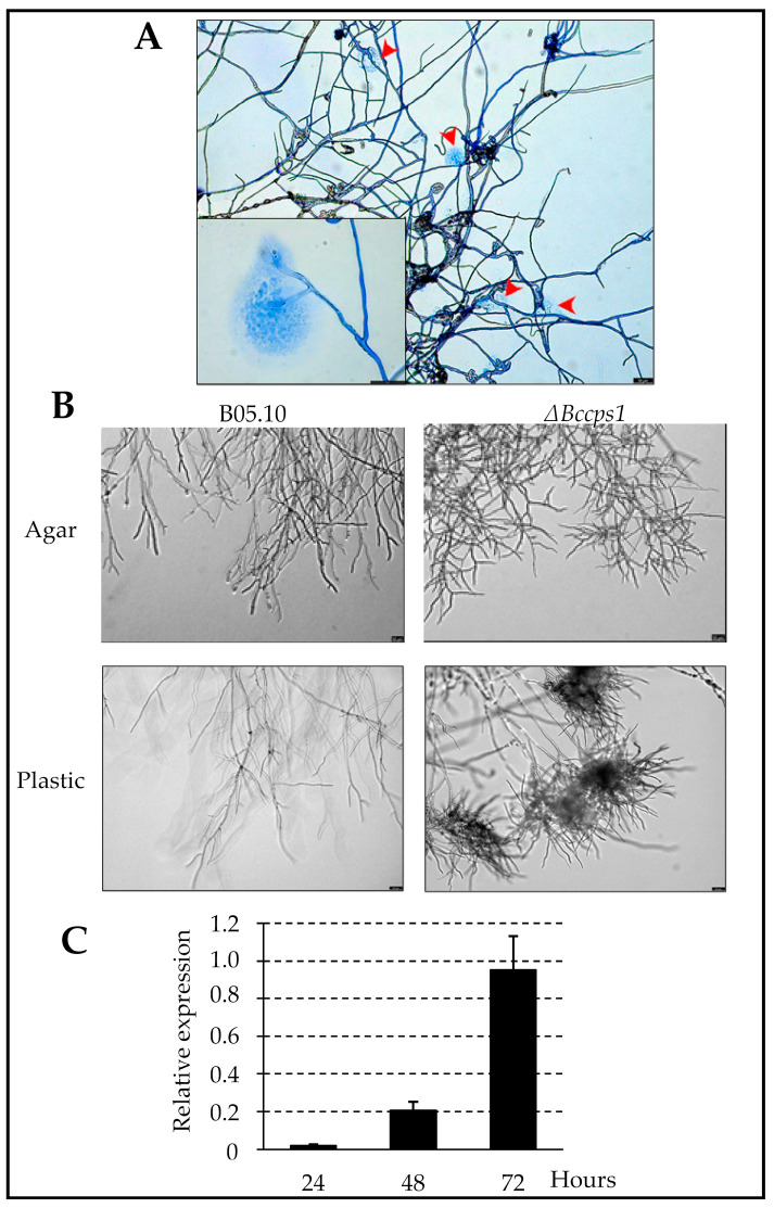
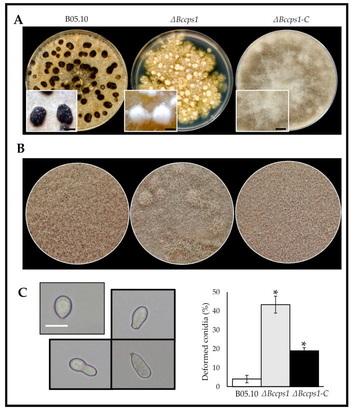
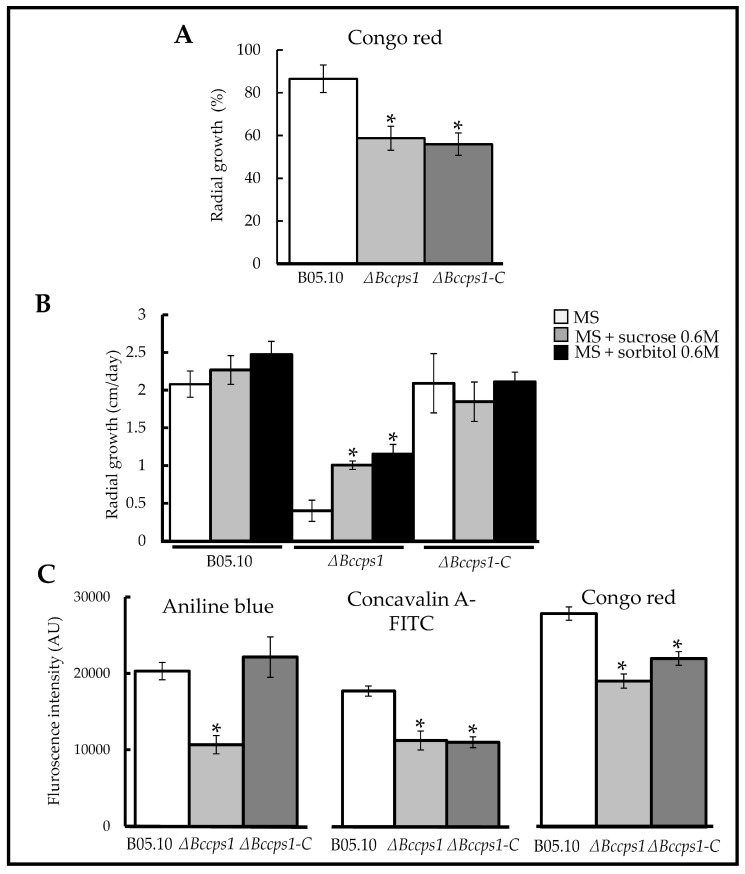
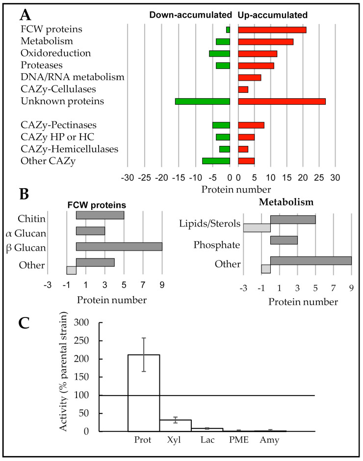
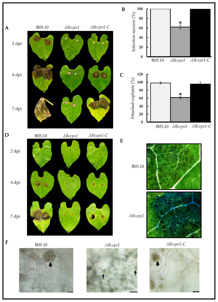
The fungal cell wall occupies a central place in the interaction between fungi and their environment. This study focuses on the role of the putative polysaccharide synthase Cps1 in the physiology, development and virulence of the grey mold-causing agent Botrytis cinerea. Deletion of the Bccps1 gene does not affect the germination of the conidia (asexual spores) or the early mycelial development, but it perturbs hyphal expansion after 24 h, revealing a two-phase hyphal development that has not been reported so far. It causes a severe reduction of mycelial growth in a solid medium and modifies hyphal aggregation into pellets in liquid cultures. It strongly impairs plant penetration, plant colonization and the formation of sclerotia (survival structures). Loss of the BcCps1 protein associates with a decrease in glucans and glycoproteins in the fungus cell wall and the up-accumulation of 132 proteins in the mutant's exoproteome, among which are fungal cell wall enzymes. This is accompanied by an increased fragility of the mutant mycelium, an increased sensitivity to some environmental stresses and a reduced adhesion to plant surface. Taken together, the results support a significant role of Cps1 in the cell wall biology of B. cinerea.

Keywords: Botrytis; adhesion; fungal cell wall; glycosyl-transferase; secretomics.


Conflict of interest statement

The authors declare no conflict of interest.

Figures

Figure 1
Figure 1
BcCps1 and its characterized homologs. (A) Presentation of cps1 genes already studied in filamentous fungi and of the Δcps1 mutant phenotypes. The simplified phylogeny is extracted from that of the Cps1 protein family (Figure S1). Membrane (Mb), Oxydative (ox). (B) Predicted secondary structure of BcCps1 inside a membrane (dotted line), showing the three TM helices, the catalytic and W domains with the conserved amino-acids (capital letters) and the non-TM hydrophobic helix (M). (C) Predicted 3D model of the B. cinerea BcCps1 protein within a lipid bilayer (blue and red tilted circles). The structure was predicted using AlphaFold Monomer v2.0 and the 3D representation was obtained using the OPM prediction tool (https://opm.phar.umich.edu/ppm_server2, 1 May 2022). The TM helices and the non-TM hydrophobic helix are colored in pink. The other protein fragments are colored following the AlphaFold confidence code (blue (high), yellow (medium), orange (low)).
Figure 2
Figure 2
BcCps1 is involved in mycelial growth. (A) Radial growth of the parental (B05.10), mutant (ΔBccps1) and complemented (ΔBccps1-C) strains on a solid rich medium over time. (B) Growth of the parental, mutant and complemented strains in a liquid rich medium. The biomass was weighed at day 3. (C) Images of the mycelial pellets collected by filtration of the liquid cultures at day 3. Black dots among the pellets are the funnel holes visible through the wet filter paper. Magnifications are shown in black circles. Scale bar: 1 cm. Stars indicate significant differences (T-test bilateral; p < 0.01).
Figure 3
Figure 3
BcCps1 is involved in hyphal development 24 h after conidia germination. (A) Rupture and cell content leakage events (red arrows) in the hyphae of the mutant strain growing out of medium droplets on a plastic surface. Hyphae were stained with cotton blue. Scale bar: 50 nm—inlet: higher magnification. (B) Parental (B05.10) and mutant (ΔBccps1) hyphal development following conidia germination (after 24 h) on a rich solid medium (Agar) and inside a droplet of rich medium on a plastic surface (Plastic). Scale bar: 50 nm. (C) Expression kinetic of the Bccps1 gene (relative to the expression of three reference genes (BcactA, Bcef1α and Bcpda1) in the parental strain grown for 72 h in a rich liquid medium. Means and standard deviations were calculated based on three independent experiments (n = 9).
Figure 4
Figure 4
BcCps1 is involved in sclerotia and conidia formation. (A) Sclerotial development in the parental (B05.10), mutant (ΔBccps1) and complemented (ΔBccps1-C) strains grown in darkness for 15 days. Dark sclerotia are visible at the surface of the parent mycelium covering the culture plate, while the mutant colony produces white cotton-like masses. Magnifications of both structures are shown in rectangles. Scale bar: 0.5 cm. (B) Images of sporulating plates of the parental, mutant and complemented strains grown under near-UV light. (C) Images (left) and counting (right) of the usual apiculate (top left) and deformed (black frame) conidia produced by the parental, mutant and complemented strains (Size bar = 10 µm). Means and standard deviations were calculated from three independent experiments (n = 150). Stars indicate significant differences (T-test bilateral; p < 0.01).
Figure 5
Figure 5
BcCps1 is involved in cell wall integrity and influences cell wall composition. (A) Sensitivity of mycelial growth to Congo red in the parental (B05.10), mutant (ΔBccps1) and complemented (ΔBccps1-C) strains. 100% corresponds to a radial growth of 2.2 cm per day. (B) Impact of 0.6 M saccharose (grey) and 0.6 M sorbitol (black) on the mycelial expansion of the B05.10, ΔBccps1 and ΔBccps1-C strains inoculated onto a rich medium (white). (C) Cell walls reactivity to anilin blue (β-1,3-glucan marker), ConcavalinA-FITC (glycoproteins marker) and Congo red (marker of chitin, some glucans and amyloid proteins) in the parental, mutant and complemented strains. Means and standard deviations were calculated from three (A,B) or six (C) independent experiments (n = 9 (A,B) or n = 17 (C)). Stars indicate significant differences (T-test bilateral; p < 0.01).
Figure 6
Figure 6
BcCps1 influences the fungal exoproteome. (A) Comparative shotgun analysis of the proteins present in the supernatants of 3-days liquid cultures of the parental and ΔBccps1 mutant strains. The proteins down- or up-accumulated in the mutant exoproteome are classified according to their functional category (fold change ≥ two, occurrence ≥ three of four biological replicates, see Table S3). CAZy, carbohydrate active enzymes; FCW, fungal cell wall; HP, hemicellulose pectin; HC, hemicellulose cellulose. (B) Sub-categorization of the categories « FCW proteins » and « Metabolism » (see Table S3). The down-accumulated proteins (green) are plotted as negative numbers. (C) Enzymatic activities measured on the same supernatants used for the proteomic analysis (four replicates). The results are shown as percentages of the parental strain values. PMEs; pectin methyl esterases. Means and standard deviations were calculated from three independent experiments (n = 12). 100% activity in the parental strain correspond to 0.41 OD280/mg dry mycelium (Proteases), 36 RFU/min.mg dry mycelium (Xylanases), 7.9 × 10⁻³ RFU/min.mg dry mycelium (Laccases), 2.35 cm/40 µL supernatant (PMEs) and 2 cm/40 µL supernatant (Amylases).
Figure 7
Figure 7
BcCps1 is involved in virulence. (A) Infection of bean leaves by mycelial explants of the parental (B05.10), mutant (ΔBccps1) and complemented (ΔBccps1-C) strains. Photos representative of the symptoms development at 2-, 4- and 7-days post inoculation (dpi) are shown. (B) Counting of the explants that succeeded at initiating infection. (C) Counting of the explants able to attach to the plant surface. Means and standard deviations were calculated from three independent experiments (n = 9). Stars indicate significant differences (T-test bilateral; p < 0.01). (D) With conidia. (E) Microscopy images of leaves collected from (D) at 2 dpi and colored with cotton blue to visualize fungal hyphae. (F) Development of infection cushions (large arrows) at 48 h of hyphal growth on plastic or glass surfaces in the parental and complemented strains. Development of hooks (thin arrows) in the mutant strain. Scale bars: 50 nm.

References

    1. Ao J., Free S.J. Genetic and biochemical characterization of the GH72 family of cell wall transglycosylases in Neurospora crassa. Fungal Genet. Biol. 2017;101:46–54. doi: 10.1016/j.fgb.2017.03.002. - DOI - PMC - PubMed
    1. Latgé J.-P. The cell wall: A carbohydrate armour for the fungal cell. Mol. Microbiol. 2007;66:279–290. doi: 10.1111/j.1365-2958.2007.05872.x. - DOI - PubMed
    1. Patel P., Free S.J. Characterization of Neurospora crassa GH16, GH17, and GH72 gene families of cell wall crosslinking enzymes. Cell Surf. 2022;8:100073. doi: 10.1016/j.tcsw.2022.100073. - DOI - PMC - PubMed
    1. Latgé J.-P., Beauvais A., Chamilos G. The Cell Wall of the Human Fungal Pathogen Aspergillus fumigatus: Biosynthesis, Organization, Immune Response, and Virulence. Annu. Rev. Microbiol. 2017;71:99–116. doi: 10.1146/annurev-micro-030117-020406. - DOI - PubMed
    1. Samar D., Kieler J.B., Klutts J.S. Identification and Deletion of Tft1, a Predicted Glycosyltransferase Necessary for Cell Wall β-1,3;1,4-Glucan Synthesis in Aspergillus fumigatus. PLoS ONE. 2015;10:e0117336. doi: 10.1371/journal.pone.0117336. - DOI - PMC - PubMed