. 2022 Aug 28;8(9):912.
doi: 10.3390/jof8090912.

Deep Learning Assisted Diagnosis of Onychomycosis on Whole-Slide Images

Affiliations

Deep Learning Assisted Diagnosis of Onychomycosis on Whole-Slide Images

Philipp Jansen et al. J Fungi (Basel). .

Abstract

Background: Onychomycosis numbers among the most common fungal infections in humans affecting finger- or toenails. Histology remains a frequently applied screening technique to diagnose onychomycosis. Screening slides for fungal elements can be time-consuming for pathologists, and sensitivity in cases with low amounts of fungi remains a concern. Convolutional neural networks (CNNs) have revolutionized image classification in recent years. The goal of our project was to evaluate if a U-NET-based segmentation approach as a subcategory of CNNs can be applied to detect fungal elements on digitized histologic sections of human nail specimens and to compare it with the performance of 11 board-certified dermatopathologists.

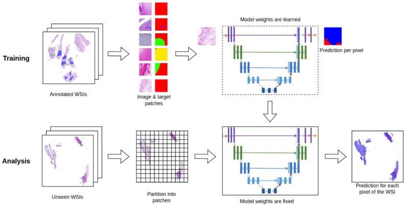
Methods: In total, 664 corresponding H&E- and PAS-stained histologic whole-slide images (WSIs) of human nail plates from four different laboratories were digitized. Histologic structures were manually annotated. A U-NET image segmentation model was trained for binary segmentation on the dataset generated by annotated slides.

Results: The U-NET algorithm detected 90.5% of WSIs with fungi, demonstrating a comparable sensitivity with that of the 11 board-certified dermatopathologists (sensitivity of 89.2%).

Conclusions: Our results demonstrate that machine-learning-based algorithms applied to real-world clinical cases can produce comparable sensitivities to human pathologists. Our established U-NET may be used as a supportive diagnostic tool to preselect possible slides with fungal elements. Slides where fungal elements are indicated by our U-NET should be reevaluated by the pathologist to confirm or refute the diagnosis of onychomycosis.

Keywords: U-NET; artificial intelligence; deep learning; dermatology; onychomycosis.


Conflict of interest statement

The authors declare no conflict of interest.

Figures

Figure 1
Figure 1
Scheme of the U-NET deep-learning architecture. Demonstrated is the workflow of training our model and using it for analysis. To train our model, we start with WSIs with sparse polygon annotations created by dermatopathologists. The annotations are processed into image and target examples of constant size. These examples are used to train our U-NET segmentation model, which learns to predict either ‘Tinea" or "Not Tinea" for every pixel in the input patch. During this process, the weights of the model are adjusted to improve predictions on training data. To use our model for analysis, we split a WSI into patches and serve each patch to the model to get a prediction. In this process, the model weights are fixed because it is not learning anymore. This also means that the model will give the same prediction for the same input patch. The predicted patches are stitched back together into an image of the same size as the original WSI, so they can be overlaid and used by pathologists to aid them in their diagnosis.
Figure 2
Figure 2
Detection of tinea on whole-slide images. Demonstrated are examples of correctly detected tinea on whole-slide images. Left: an overview of the entire nail section processed; right: zoomed-in examples showing the PAS-stained tinea elements and calls by the algorithm detecting tinea elements, with red pixels representing Tinea. Blurry area on the top image shows areas of the slide where the nail material was not in focus.
Figure 3
Figure 3
Examples of false-positive tinea calls. Examples where the algorithm made a call of tinea but was not verified by the majority of dermatopathologists are shown. The left column represents PAS-stained slides; the right picture with a heatmap overlay of the U-NET algorithm. The high amount of staining artifacts, common for difficult-to-process nail material, is apparent, which poses a diagnostic difficulty for pathologists and the algorithm alike.
Figure 4
Figure 4
Performance of pathologists and U-NET. Accuracy of each pathologist is depicted in blue. Accuracy of our model is depicted in purple. Correct classification for each case was determined by majority voting across pathologists' representation.
Figure 5
Figure 5
ROC curve comparing 11 dermatopathologists with U-NET algorithm. Demonstrated are individual dermatopathologists as well as U-NET algorithm in terms of sensitivity (true positive rate, recall) and 1-specificity (false positive rate). In the presented plot, cases where pathologists made a "maybe" call were not considered for their performance.

References

    1. Gupta A.K., Taborda V.B.A., Taborda P.R.O., Shemer A., Summerbell R.C., Nakrieko K.A. High prevalence of mixed infections in global onychomycosis. PLoS ONE. 2020;15:e0239648. doi: 10.1371/journal.pone.0239648. - DOI - PMC - PubMed
    1. Gupta A.K., Versteeg S.G., Shear N.H. Onychomycosis in the 21st Century: An Update on Diagnosis, Epidemiology, and Treatment. J. Cutan. Med. Surg. 2017;21:525–539. doi: 10.1177/1203475417716362. - DOI - PubMed
    1. Guibal F., Baran R., Duhard E., de Chauvin M.F. Epidemiology and management of onychomycosis in private dermatological practice in France. Ann. Dermatol. Venereol. 2008;135:561–566. doi: 10.1016/j.annder.2008.05.004. - DOI - PubMed
    1. Koshnick R.L., Lilly K.K., St Clair K., Finnegan M.T., Warshaw E.M. Use of diagnostic tests by dermatologists, podiatrists and family practitioners in the United States: Pilot data from a cross-sectional survey. Mycoses. 2007;50:463–469. doi: 10.1111/j.1439-0507.2007.01422.x. - DOI - PubMed
    1. Wollina U., Nenoff P., Haroske G., Haenssle H.A. The Diagnosis and Treatment of Nail Disorders. Dtsch. Arztebl. Int. 2016;113:509–518. doi: 10.3238/arztebl.2016.0509. - DOI - PMC - PubMed

LinkOut - more resources