. 2022 Sep 19;8(9):979.
doi: 10.3390/jof8090979.

Biodiversity and Bioprospecting of Fungal Endophytes from the Antarctic Plant Colobanthus quitensis

Affiliations

Biodiversity and Bioprospecting of Fungal Endophytes from the Antarctic Plant Colobanthus quitensis

Laura Bertini et al. J Fungi (Basel). .

Abstract

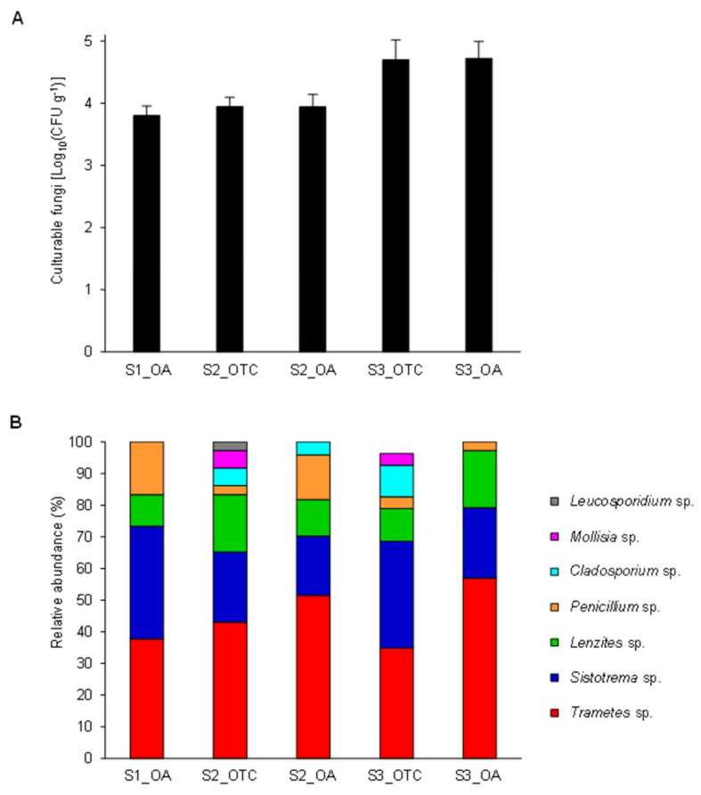
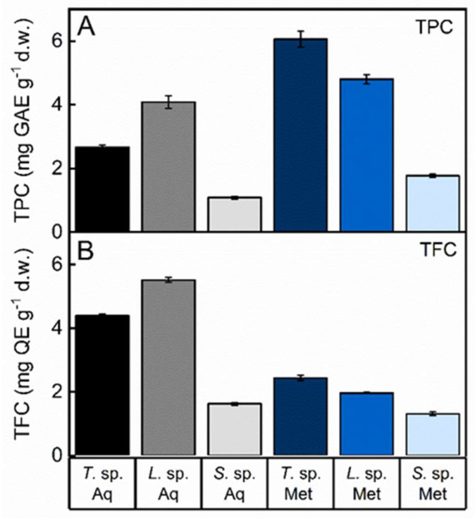
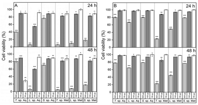
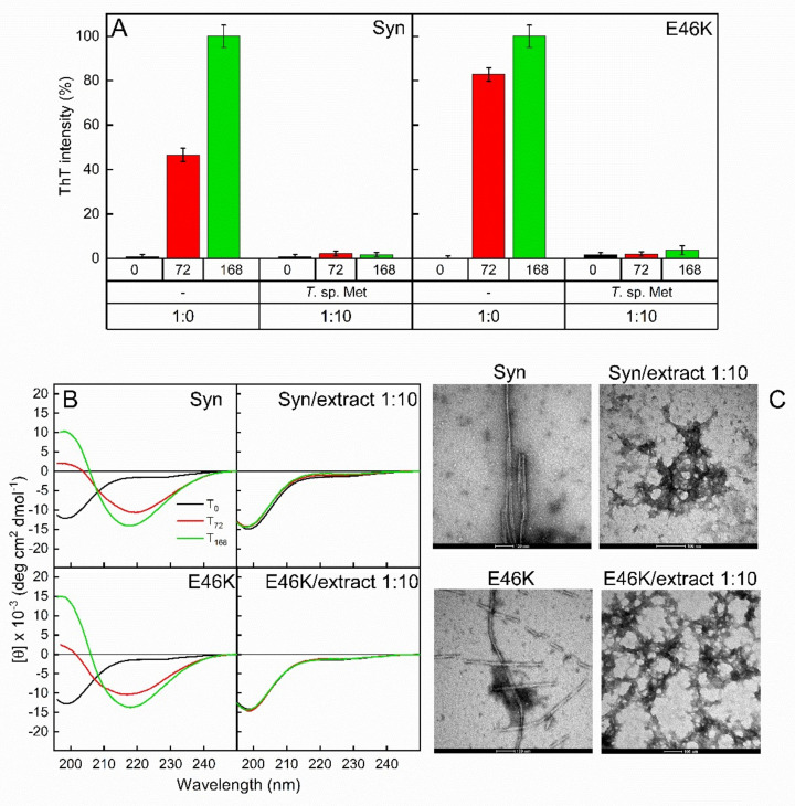
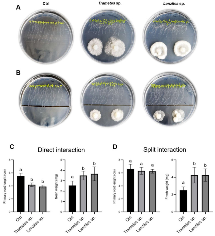
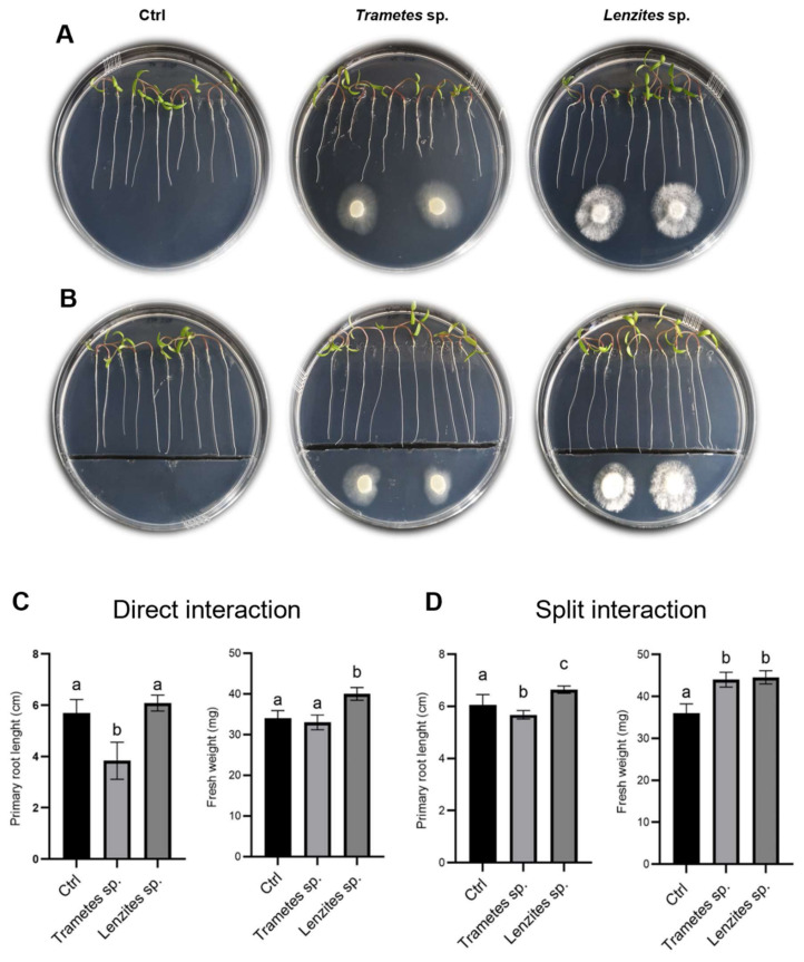
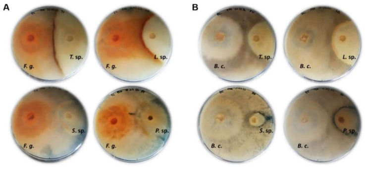
Microorganisms from extreme environments are considered as a new and valuable reservoir of bioactive molecules of biotechnological interest and are also utilized as tools for enhancing tolerance to (a)biotic stresses in crops. In this study, the fungal endophytic community associated with the leaves of the Antarctic angiosperm Colobanthus quitensis was investigated as a new source of bioactive molecules. We isolated 132 fungal strains and taxonomically annotated 26 representative isolates, which mainly belonged to the Basidiomycota division. Selected isolates of Trametes sp., Lenzites sp., Sistotrema sp., and Peniophora sp. displayed broad extracellular enzymatic profiles; fungal extracts from some of them showed dose-dependent antitumor activity and inhibited the formation of amyloid fibrils of α-synuclein and its pathological mutant E46K. Selected fungal isolates were also able to promote secondary root development and fresh weight increase in Arabidopsis and tomato and antagonize the growth of pathogenic fungi harmful to crops. This study emphasizes the ecological and biotechnological relevance of fungi from the Antarctic ecosystem and provides clues to the bioprospecting of Antarctic Basidiomycetes fungi for industrial, agricultural, and medical applications.

Keywords: Antarctic fungi; DNA barcoding; bioactive compounds; culturomics; extracellular enzymes; plant–fungus interaction.


Conflict of interest statement

The authors declare no conflict of interest.

Figures

Figure 1
Figure 1
Endophytic fungi from Colobanthus quitensis leaves. (A) Colony-forming units (CFU) of culturable endophytic fungi per fresh plant weight unit (g) were evaluated for C. quitensis plantlets harvested in open areas (OA samples) as well as inside open-top chambers (OTC samples). Culturable fungi were grown at 25 ± 1 °C for 21 days on potato dextrose agar (PDA) supplemented with 0.25% lactic acid. No additional colonies could be observed for a longer incubation time. Mean Log10 (CFU g−1) and standard deviation values from three replicates (each as a pool of plants) are presented for each sample. (B) Relative abundances of taxa of culturable endophytic fungi assessed by morphological analyses. No significant differences were found among the collection sites (OA and OTC), according to Tukey’s test (p > 0.05).
Figure 2
Figure 2
Total phenolic content (TPC) (A) and total flavonoid content (TFC) (B) of the aqueous (Aq) and methanol (Met) fungal extracts of Trametes sp. S2.OA.C_F6 (T. sp.), Lenzites sp. S3.OA.B_F6 (L. sp.) and Sistotrema sp. S1.OA.C_F2 (S. sp.). TPC was expressed in terms of gallic acid equivalent (GAE) g−1 dry weight; TFC was expressed as quercetin equivalent (QE) g−1 dry weight. Data were reported as mean values ± standard deviation (SD) of triplicates.
Figure 3
Figure 3
Cell viability test. SH-SY5Y (A) and HBEC (B) cells exposed for 24 and 48 h to increasing concentrations (50, 100, 500 µg) of aqueous (Aq) or methanol (Met) fungal extracts of Trametes sp. S2.OA.C_F6 (T. sp.), Lenzites sp. S3.OA.B_F6 (L. sp.) and Sistotrema sp. S1.OA.C_F2 (S. sp.). Results were expressed as a percentage of the corresponding untreated cultures. Error bars indicate the standard error of three independent experiments carried out in triplicate. Asterisks indicate statistically significant differences: *, p < 0.05; **, p < 0.01; ***, p < 0.001.
Figure 4
Figure 4
Amyloid formation inhibition probed by ThT fluorescence assay (A), circular dichroism (CD) (B) and TEM analysis (C). α-Synuclein (Syn) and its familial mutant (E46K) were left to aggregate in the absence (1:0) and in the presence (1:10) of methanol extract of Trametes sp. S2.OA.C_F6 (T. sp. Met). Aliquots taken from the mixtures were collected after 0 (black), 72 (red) and 168 h (green) of incubation. The TEM scale bar in every picture corresponds to 100 nm. Error bars indicate the standard error of three independent experiments carried out in triplicate.
Figure 5
Figure 5
Effect of Trametes sp. S2.OA.C_F6 and Lenzites sp. S3.OA.B_F6 isolates on Arabidopsis growth. (A,B) Representative photos of 12-day-old Arabidopsis (Col-0) plantlets grown on MS½ medium at 7 dpi. (A) Direct interaction experiments and (B) split interaction experiments. (C,D) Physiological parameter measurements. Primary root length (cm) and fresh weight (mg) measured in direct interaction experiments and (C) split interaction experiments (D). Bar plots represent mean ± SD (n = 15). Different letters indicate significant differences (one-way ANOVA, p < 0.05; Tukey post hoc test, p < 0.05).
Figure 6
Figure 6
Effect of Trametes sp. S2.OA.C_F6 and Lenzites sp. S3.OA.B_F6 isolates on tomato growth. (A,B) Representative photos of 8-day-old tomato plantlets grown on MS½ medium at 3 dpi. (A) Direct interaction experiments and (B) split interaction experiments. (C,D) Physiological parameter measurements. Primary root length (cm) and fresh weight (mg) measured in direct interaction experiments (C) and split interaction experiments (D). Bar plots represent mean ± SD (n = 15). Different letters indicate significant differences (one-way ANOVA, p < 0.05; Tukey post hoc test, p < 0.05).
Figure 7
Figure 7
Co-cultivation of Fusarium graminearum (A) and Botrytis cinerea (B) (on the left of the plates) and Antarctic fungal isolates on PDA solid medium at 10 dpi. F. g.: Fusarium graminearum; B. c.: Botrytis cinerea; T. sp.: Trametes sp.; L. sp.: Lenzites sp.; S. sp.: Sistotrema sp.; P. sp.: Peniophora sp.

References

    1. Danilovich M.E., Sánchez L.A., Acosta F.F., Delgado O.D. Antarctic bioprospecting: In pursuit of microorganisms producing new antimicrobials and enzymes. Polar Biol. 2018;41:1417–1433. doi: 10.1007/s00300-018-2295-4. - DOI
    1. Raddadi N., Cherif A., Daffonchio D., Neifar M., Fava F. Biotechnological applications of extremophiles, extremozymes and extremolytes. Appl. Microbiol. Biotechnol. 2015;99:7907–7913. doi: 10.1007/s00253-015-6874-9. - DOI - PubMed
    1. Timling I., Taylor D.L. Peeking through a frosty window: Molecular insights into the ecology of Arctic soil fungi. Fungal Ecol. 2012;5:419–429. doi: 10.1016/j.funeco.2012.01.009. - DOI
    1. Weinstein R.N., Montiel P.O., Johnstone K. Influence of growth temperature on lipid and soluble carbohydrate synthesis by fungi isolated from fellfield soil in the maritime Antarctic. Mycologia. 2000;92:222–229. doi: 10.1080/00275514.2000.12061148. - DOI
    1. Coleine C., Stajich J.E., de los Ríos A., Selbmann L. Beyond the extremes: Rocks as ultimate refuge for fungi in drylands. Mycologia. 2021;113:108–133. doi: 10.1080/00275514.2020.1816761. - DOI - PubMed

LinkOut - more resources